. 2024 Dec;28(23):e70270.
doi: 10.1111/jcmm.70270.

Long Non-Coding RNA LINC01116 Promotes the Proliferation of Lung Adenocarcinoma by Targeting miR-9-5p/CCNE1 Axis

Affiliations

Long Non-Coding RNA LINC01116 Promotes the Proliferation of Lung Adenocarcinoma by Targeting miR-9-5p/CCNE1 Axis

Hui Zhang et al. J Cell Mol Med. 2024 Dec.

Abstract

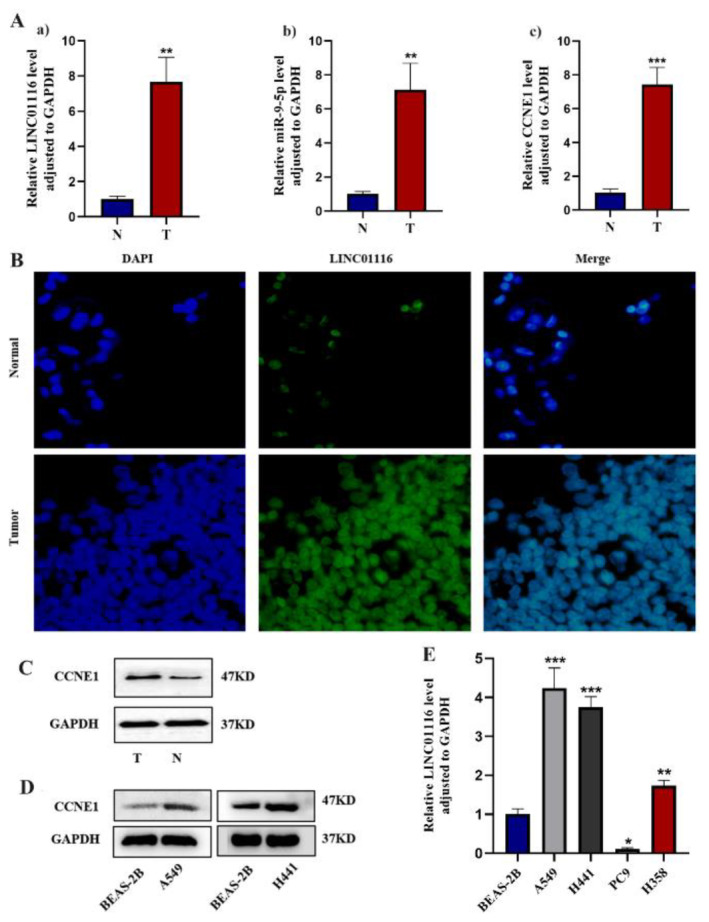
Long non-coding RNA (lncRNA) LINC01116 is crucial in promoting cell proliferation, invasion and migration in solid tumours, including lung adenocarcinoma (LUAD). LINC01116 acts as a competing endogenous RNAs (ceRNA) that binds competitively to microRNAs and plays a critical role in tumour migration and invasion. However, other mechanisms of action besides the ceRNA theory have been rarely reported and remain to be elucidated further. The differences in RNA and protein levels in cells and tissues were assessed through real-time quantitative PCR and Western blot analysis. In vitro functional assays and in vivo xenograft models were used to analyse the function of LINC01116 in LUAD. Thus, the molecular correlation between miR-9-5p and CCNE1 was investigated through direct and indirect mechanism experiments. LINC01116, miR-9-5p and CCNE1 were upregulated in LUAD cell lines and tissues and were associated with a poor prognosis in patients. LINC01116 depletion inhibited proliferation but facilitated cell apoptosis. AGO2-RNA binding protein immunoprecipitation (AGO2-RIP) experiments confirmed that AGO2 binds to LINC01116 and miR-9-5p, indicating that LINC01116 interacts with miR-9-5p. The overexpression of miR-9-5p and CCNE1 effectively counteracts the biological effects of LINC01116 knockdown on reduced proliferation and cell cycle arrest in LUAD cells. The downregulation of miR-9-5p significantly reduces the CCNE1 level in A549 cells, and the upregulation of LINC01116 counteracts the downregulation of miR-9-5p effect, restoring the expression level of CCNE1. Our data demonstrated that LINC01116 regulates the expression of CCNE1 by positively regulating miR-9-5p, thereby affecting cell cycle, proliferation and participating in the development of LUAD.

Keywords: CCNE1; LINC01116; LUAD; cell proliferation; miR‐9‐5p.


Conflict of interest statement

The authors declare no conflicts of interest.

Figures

FIGURE 1
FIGURE 1
(A) The ceRNA network of the co‐expression of lncRNAs, miRNAs and mRNAs was obtained by WGCNA analysis. (B) Expression of LINC01116 in LUAD and normal tissue samples. The expression level of LINC01116 in LUAD samples was significantly higher than that in normal tissues (***p < 0.001). (C) Kaplan–Meier survival analysis was applied to show significantly different survival curves between LUAD patients with high and low expression levels of LINC01116 (p = 0.0019). The abscissa of the survival curve is the observation time, and the ordinate is the overall survival rate. (D. a) Hypothetical binding site of LINC01116 and miR‐9‐5p. (b). starBase database predicted the positive correlation between LINC01116 and miR‐9‐5p (p = 3.38e‐06). (E. a) Hypothetical binding site of miR‐9‐5p and CCNE1. (b) starBase database predicted the positive correlation between LINC01116 and miR‐9‐5p (p = 1.97e‐17). (F) starBase database predicted the positive correlation between LINC01116 and CCNE1 (p = 0.012).
FIGURE 2
FIGURE 2
(A) The expression level of LINC01116, miR‐9‐5p and CCNE1 in tumour tissue (T) was analysed by qRT‐PCR [**p < 0.01, ***p < 0.001 vs. normal tissue (N)]. (B) Sub‐cellular localization of LINC01116 by FISH assays and its statistical analysis (**p < 0.01). (C) Protein expression of CCNE1 in tumour tissue measured by the Western blot analysis (*p < 0.05 vs. normal tissue). (D) Protein expression of CCNE1 in A549 or H441 cells measured by the Western blot analysis. (E) Expression of LINC01116 in various cell lines was quantitatively assessed via qRT‐PCR (*p < 0.05, **p < 0.01, ****p < 0.0001 vs. BEAS‐2B).
FIGURE 3
FIGURE 3
(A: a). Silencing efficiency of sh‐LINC01116 in A549 cells (**p < 0.01). (b and c). Relative expression level of miR‐9‐5p and CCNE1 in the sh‐LINC01116 group was assessed by qRT‐PCR analysis (*p < 0.05, **p < 0.01, ***p < 0.001 vs. NC group). (B) CCK‐8 assay showed that LINC01116 stable knockdown inhibited the proliferation of A549 cells. (C) Protein levels of CCNE1, CDK2, Ki‐67, MCM7, PCNA, p53 and p16 in the sh‐LINC01116 group, as measured by the Western blot. (D) Apoptosis rate of A549 cells, treated with negative control and LINC01116 shRNA (**p < 0.01). (E) Cell cycle of A549 cells, treated with negative control and LINC01116 shRNA, was analysed by flow cytometry (***p < 0.001).
FIGURE 4
FIGURE 4
(A) CCK‐8 assay showed that LINC01116 stable knockdown inhibited the growth of A549 cells, and the overexpression of miR‐9‐5p or CCNE1 promoted A549 cell growth (***p < 0.001). (B) EdU assays were employed to assess the cell proliferation capacity after the downregulation of LINC01116 and upregulation of miR‐9‐5p or CCNE1. (C) After treatment with negative control, si‐LINC01116 or pcDNA3.1‐miR‐9‐5p/pcDNA3.1‐CCNE1 plasmid, A549 cells were stained and analysed by flow cytometry (***p < 0.001). (D) Protein expression of CCNE1, CDK2, Ki‐67, MCM7, PCNA, p53 and p16 in the si‐LINC01116 and pcDNA3.1‐miR‐9‐5p groups or pcDNA3.1‐CCNE1 group, as measured by the Western blot.
FIGURE 5
FIGURE 5
(A) qRT‐PCR based on RIP indicated that both LINC01116 and miR‐9‐5p can bind to miR‐744‐5p. One‐way ANOVA was applied for analysis (**p < 0.01 ***p < 0.001). (B) Protein level of CCNE1, regulated by LINC01116, was measured by the Western blot analysis. (C) Protein level of CCNE1, regulated by miR‐9‐5p, was estimated by the Western blot analysis.
FIGURE 6
FIGURE 6
(A) The right panels: cells were transplanted with sh‐NC, sh‐LINC01116, pcDNA3.1‐ miR‐9‐5p or CCNE1 plasmids (n = 3). The left panels: the weight and volume of each tumour were measured (*p < 0.05, **p < 0.01, ***p < 0.001). (B) The tumour weight in the sh‐LINC01116 group was decreased, and the pcDNA3.1‐miR‐9‐5p/CCNE1 group was increased (*p < 0.05, **p < 0.01, ***p < 0.001). (C) The tumour volume was decreased in the sh‐LINC01116 group but increased in the pcDNA3.1‐miR‐9‐5p/CCNE1 group (*p < 0.05, **p < 0.01, ***p < 0.001). (D and E) The relative expression level of LINC01116 and miR‐9‐5p in the tumour tissue of BALB/C nude mice transplanted with sh‐NC, sh‐LINC01116, pcDNA3.1‐miR‐9‐5p or CCNE1 plasmids was achieved by qRT‐PCR (*p < 0.05, ***p < 0.001). (F) Protein expression of CCNE1, CDK2, Ki‐67 MCM7, PCNA and p16 in the tumour tissue of BALB/C nude mice, as measured by the Western blot analysis.
FIGURE 7
FIGURE 7
Kaplan–Meier survival analysis shows significantly different survival curves between LUAD patients with high and low expression levels of CCNE1 (p = 0.0058). The abscissa of the survival curve is the observation time, and the ordinate is the overall survival rate.

Similar articles

Cited by

References

    1. Oudkerk M., Liu S. Y., Heuvelmans M. A., Walter J. E., and Field J. K., “Lung Cancer LDCT Screening and Mortality Reduction – Evidence, Pitfalls and Future Perspectives,” Nature Reviews. Clinical Oncology 18, no. 3 (2021): 135–151, 10.1038/s41571-020-00432-6. - DOI - PubMed
    1. Chen Z., Fillmore C. M., Hammerman P. S., Kim C. F., and Wong K. K., “Non‐small‐Cell Lung Cancers: A Heterogeneous Set of Diseases,” Nature Reviews. Cancer 14, no. 8 (2014): 535–546, 10.1038/nrc3775. - DOI - PMC - PubMed
    1. Balzer B. W. R., Loo C., Lewis C. R., Trahair T. N., and Anazodo A. C., “Adenocarcinoma of the Lung in Childhood and Adolescence: A Systematic Review,” Journal of Thoracic Oncology 13, no. 12 (2018): 1832–1841, 10.1016/j.jtho.2018.08.2020. - DOI - PubMed
    1. Denisenko T. V., Budkevich I. N., and Zhivotovsky B., “Cell Death‐Based Treatment of Lung Adenocarcinoma,” Cell Death & Disease 9, no. 2 (2018): 117, 10.1038/s41419-017-0063-y. - DOI - PMC - PubMed
    1. Goldstraw P., Chansky K., Crowley J., et al., “The IASLC Lung Cancer Staging Project: Proposals for Revision of the TNM Stage Groupings in the Forthcoming (Eighth) Edition of the TNM Classification for Lung Cancer,” Journal of Thoracic Oncology 11, no. 1 (2016): 39–51, 10.1016/j.jtho.2015.09.009. - DOI - PubMed

MeSH terms

LinkOut - more resources