Effect of patient-contextual skin images in human- and artificial intelligence-based diagnosis of melanoma: Results from the 2020 SIIM-ISIC melanoma classification challenge
- PMID: 39648687
- PMCID: PMC12145458
- DOI: 10.1111/jdv.20479
Effect of patient-contextual skin images in human- and artificial intelligence-based diagnosis of melanoma: Results from the 2020 SIIM-ISIC melanoma classification challenge
Abstract
Background: While the high accuracy of reported AI tools for melanoma detection is promising, the lack of holistic consideration of the patient is often criticized. Along with medical history, a dermatologist would also consider intra-patient nevi patterns, such that nevi that are different from others on a given patient are treated with suspicion.
Objective: To evaluate whether patient-contextual lesion-images improves diagnostic accuracy for melanoma in a dermoscopic image-based AI competition and a human reader study.
Methods: An international online AI competition was held in 2020. The task was to classify dermoscopy images as melanoma or benign lesions. A multi-source dataset of dermoscopy images grouped by patient were provided, and additional use of public datasets was permitted. Competitors were judged on area under the receiver operating characteristic (AUROC) on a private leaderboard. Concurrently, a human reader study was hosted using a subset of the test data. Participants gave their initial diagnosis of an index case (melanoma vs. benign) and were then presented with seven additional lesion-images of that patient before giving a second prediction of the index case. Outcome measures were sensitivity and specificity.
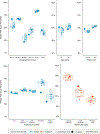
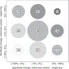
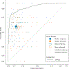
Results: The top 50 of 3308 AI competition entries achieved AUROC scores ranging from 0.943 to 0.949. Few algorithms considered intra-patient lesion patterns and instead most evaluated images independently. The median sensitivity and specificity of human readers before receiving contextual images were 60.0% and 86.7%, and after were 60.0% and 85.7%. Human and AI algorithm performance varied by image source.
Conclusions: This study provided an open-source state-of-the-art algorithm for melanoma detection that has been evaluated at multiple centres. Patient-contextual images did not positively impact performance of AI algorithms or human readers. Providing seven contextual images and no total body image may have been insufficient to test the applicability of the intra-patient lesion patterns.
© 2024 European Academy of Dermatology and Venereology.
Conflict of interest statement
CONFLICT OF INTEREST STATEMENT
HPS is a shareholder of MoleMap NZ Limited and e-derm consult GmbH and undertakes regular teledermatological reporting for both companies. HPS is a Medical Consultant for Canfield Scientific Inc. and a Medical Advisor for First Derm. HPS received a NHMRC Synergy Grant (2009923) and receives research funding from the Australian Cancer Research Foundation and NHMRC Centre of Research Excellence (2006551). VR is funded by the Melanoma Research Alliance, has a contract with Lutris Pharma, receives research support from Kaggle and AWS, receives consulting fees and stock options from Inhabit Brands Inc. and sits on both the AAD AUI committee and SIIM Board. AH receives consulting fees from Canfield Scientific Inc. and SciBase, participates on an advisory board for Jannsen, participates in the Organizing Committee of the International Skin Imaging Society, is Vice President of the Skin Cancer Foundation and is co-founder of SpotDoc. NRK, AH, JW and VR receive research funding through the MSKCC Cancer Center Support Grant P30 CA008748. BBS anticipates employment with Canfield Scientific, Inc. PG has received honoraria from MetaOptima and TPY. JK was awarded a grant from the Melanoma Research Alliance, received consulting fees from The Skin Diary Ltd. and SharkNinja LLC, received personal payment from Beiersdorf LTD and Leo Pharmaceuticals LTD, and received travel support from Almirall Pharmaceuticals LTD. HK received royalties from CASIO, Heine and MetaOptima; received consulting fees from FotoFinder and AI Medical Technology; received payment or honoraria from FotoFinder, Pelpharma, La Roche Posay, Eli Lilly, Novartis and MSD; holds a leadership position at the International Dermoscopy Society; and received equipment from FotoFinder, Heine, and CASIO. JP is co-founder of Athena Tech, a scientific advisor of Dermavision and chairman of the EADV Task Force of Artificial Intelligence. PT received a grant/contract from Lilly; payment/honoraria for lectures/presentations from FotoFinder, Novartis, Lilly and AbbVie; and holds a leadership position at the International Dermoscopy Society. CAP, MC, KL and CR have no COI to report.
Figures


References
-
- Tschandl P, Rinner C, Apalla Z, Argenziano G, Codella N, Halpern A, et al. Human-computer collaboration for skin cancer recognition. Nat Med. 2020;26(8):1229–34. - PubMed
-
- Cerminara SE, Cheng P, Kostner L, Huber S, Kunz M, Maul JT, et al. Diagnostic performance of augmented intelligence with 2D and 3D total body photography and convolutional neural networks in a high-risk population for melanoma under real-world conditions: a new era of skin cancer screening? Eur J Cancer. 2023;190:112954. - PubMed
MeSH terms
Grants and funding
LinkOut - more resources
Full Text Sources
Medical

