. 2025 Dec;57(1):2437046.
doi: 10.1080/07853890.2024.2437046. Epub 2024 Dec 9.

Development and application of an uncapped mRNA platform

Affiliations

Development and application of an uncapped mRNA platform

Xiaodi Zheng et al. Ann Med. 2025 Dec.

Abstract

Background: A novel uncapped mRNA platform was developed.

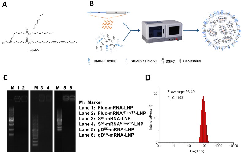
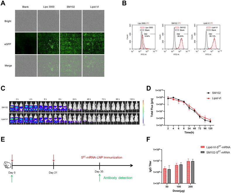
Methods: Five lipid nanoparticle (LNP)-encapsulated mRNA constructs were made to evaluate several aspects of our platform, including transfection efficiency and durability in vitro and in vivo and the activation of humoral and cellular immunity in several animal models. The constructs were eGFP-mRNA-LNP (for enhanced green fluorescence mRNA), Fluc-mRNA-LNP (for firefly luciferase mRNA), SδT-mRNA-LNP (for Delta strain SARS-CoV-2 spike protein trimer mRNA), gDED-mRNA-LNP (for truncated glycoprotein D mRNA coding ectodomain from herpes simplex virus type 2 (HSV2)) and gDFR-mRNA-LNP (for truncated HSV2 glycoprotein D mRNA coding amino acids 1-400).

Results: Quantifiable target protein expression was achieved in vitro and in vivo with eGFP- and Fluc-mRNA-LNP. SδT-mRNA-LNP, gDED-mRNA-LNP and gDFR-mRNA-LNP induced both humoral and cellular immune responses comparable to those obtained by previously reported capped mRNA-LNP constructs. Notably, SδT-mRNA-LNP elicited neutralizing antibodies in hamsters against the Omicron and Delta strains. Additionally, gDED-mRNA-LNP and gDFR-mRNA-LNP induced potent neutralizing antibodies in rabbits and mice. The mRNA constructs with uridine triphosphate (UTP) outperformed those with N1-methylpseudouridine triphosphate (N1mψTP) in the induction of antibodies via SδT-mRNA-LNP.

Conclusions: Our uncapped, process-simplified and economical mRNA platform may have broad utility in vaccines and protein replacement drugs.KEY MESSAGESThe mRNA platform described in our paper uses internal ribosome entry site (IRES) (Rapid, Amplified, Capless and Economical, RACE; Register as BH-RACE platform) instead of caps and uridine triphosphate (UTP) instead of N1-methylpseudouridine triphosphate (N1mψTP) to synthesize mRNA.Through the self-developed packaging instrument and lipid nanoparticle (LNP) delivery system, mRNA can be expressed in cells more efficiently, quickly and economically.Particularly exciting is that potent neutralizing antibodies against Delta and Omicron real viruses were induced with the new coronavirus S protein mRNA vaccine from the BH-RACE platform.

Keywords: LNP; Omicron strain neutralization; Uncapped mRNA.


Conflict of interest statement

Peng Ni, Linkang Cai, Xiaotai Shi, Zonghuang Ke, Siqi Zhang, Binfeng Yang, Wei Long, Zhong Wang, Zhizheng Fang, Yan Xu, Hanming Wang and Binlei Liu are employed by Wuhan Binhui Biopharmaceutical Co., Ltd. No potential conflict of interest was reported by the author(s).

Figures

Figure 1.
Figure 1.
Amplification of the IVT template and preparation of uncapped mRNA with high yield and purity. (A) Schematic diagram of the uncapped mRNA structures. (B) Scatter plot of IVT template concentrations obtained by PCR amplification. Data are presented as the mean ± SD (n = 3). (C) Agarose gel (1%) electropherogram of IVT-synthesized mRNA. Lanes 1–7 were the 7-repeat IVT products, the sample loading volume was 1 μL, and all IVT templates were Fluc mRNA; M was a 2000 bp DNA marker with a loading volume of 5 μL; lanes 8, 9 and 10 were 900, 500 and 250 ng RNA references, respectively. (D) SEC-HPLC analysis of mRNA samples obtained before and after purification. Red and blue diagrams represent pre- and post-purified mRNA samples. The major peak area of the purified sample was greater than 99%.
Figure 2.
Figure 2.
Encapsulation of mRNA. (A) The structure of Lipid-VI. (B) Process flow diagram. (C) mRNA-LNP electrophoresis with parameters of 1% agarose gel, 120 V and TAE running buffer. M: marker; lane 1: Fluc-mRNA-LNP; lane 2: FlucN1mψTP-mRNA-LNP; lane 3: SδT-mRNA-LNP; lane 4: SδT-mRNAN1mψTP-LNP; lane 5: gDED-mRNA-LNP; lane 6: gDFR-mRNA-LNP. (D) Typical particle size of mRNA-LNP detected with a mastersizer laser diffraction particle size analyser.
Figure 3.
Figure 3.
Uncapped mRNA-LNP transfection in vivo. (A) The live luciferase fluorescence images of mice at various time points after treated with different mRNA-LNP. (B) Quantification of the bioluminescent signal measured in (A). Means and SEM are shown (n = 6). (C) The luciferase fluorescence images of mice treated with different doses of mRNA-LNP at 4 h post-transfection. (D) Quantification of the bioluminescent signal measured in (C). Means and SEM are shown (n = 3). The cap UTP means that the mRNA has a 5′ cap and prepared with uridine triphosphate. The cap N1mψTP means that the mRNA has a 5′ cap and prepared with N1-methyl pseudo uridine triphosphate. The uncapped UTP means the mRNA does not have a 5′ cap and prepared with uridine triphosphate. Significance was calculated using Student’s t-test (*p < .05; **p < .01).
Figure 4.
Figure 4.
Evaluation of the transfection and the immunogenicity of mRNA encapsulated with Lipid-VI. (A) Bright-field and dark-field photos of BHK cells transfected with eGFP-mRNA-LNP for 24 h. (B) The transfection efficiency was detected by flow cytometry. (C) The luciferase fluorescence images of mice intramuscularly injected with 30 µg of Fluc mRNA encapsulated in SM102 and Lipid-VI. (D) Quantification of the bioluminescent signal measured in (C). Means and SEM are shown (n = 3). (E) Schematic diagram of immunization and sample collection in Syrian hamsters. (F) The SARS-CoV-2-specific IgG antibody titers of the SδT-mRNA encapsulated with SM102 and Lipid-VI on day 35 were determined by ELISA. Means and SEM are shown (n = 3).
Figure 5.
Figure 5.
Antibody detection after immunization with SδT-mRNA-LNP and SδT-mRNAN1mψTP-LNP. (A) Schematic diagram of immunization and serum sample collection in C57BL/6 mice. (B) The SARS-CoV-2-specific IgG antibody titers of the three groups (0 µg, 30 μg N1mψTP and 30 μg UTP) were determined by ELISA. Means and SEM are shown (n = 3). Significance was calculated using Student’s t-test (**p < .01). (C) Schematic diagram of immunization and sample collection in Syrian hamsters. (D) The SARS-CoV-2-specific IgG antibody titers of the four groups on day 35 were determined by ELISA. Means and SEM are shown (n = 3).
Figure 6.
Figure 6.
Detection of the neutralizing antibodies produced by animals immunized with gDED-mRNA-LNP and gDFR-mRNA-LNP using Vero cells and virus-infected plaques. (A) Schematic diagram of immunization and serum sample collection. The red and green arrows represent the immunization time and the blood collection time, respectively. (B) The postimmunization serum (at day 56) neutralization of oHSV2-eGFP. DME/F-12 containing neonatal bovine serum (termed medium), preimmunization serum (termed serum-naïve), postimmunization serum from a mouse immunized with gDFR-mRNA-LNP (termed serum-gDFR) and postimmunization serum from a mouse immunized with gDED-mRNA-LNP (termed serum-gDED) were incubated with oHSV2-eGFP. (C) Rabbit immunization regimen of gDED-mRNA-LNP. Three rabbits received two doses of gDED-mRNA-LNP immunization on days 0 and 14 (one via intradermal injection, two via intramuscular). Blood was collected on days 0, 14 and 28 to separate the serum for the neutralization assay. (D) The serum neutralization antibody titers (on day 28) were determined using Vero cells and plaques observed 72 h post-virus infection for: (a) the 20 μg intradermal injection group with serum diluted 64 times; (b) the 20 μg intradermal injection group with serum diluted 512 times; (c) the 25 μg intramuscular injection group with serum diluted 16 times; (d) the 50 μg intramuscular injection group with serum diluted 32 times; and (e) the 50 μg intramuscular injection group with serum diluted 64 times. The constant amount of virus was 100 CCID50 per well, and the serum (collected at 28 days) was diluted for neutralizing antibody detection.
Figure 7.
Figure 7.
Animal T-cell immune responses after gDED-mRNA-LNP/gDFR-mRNA-LNP or SδT-mRNA-LNP immunization. (A, D) Schematic diagram of immunization and serum sample collection. The red and green arrows represent the immunization time and detection time, respectively. (B, E) The number of spots formed by IFN-γ cytokines secreted by splenocytes after immunization. (C, F) Quantification of spot formation by IFN-γ cytokines secreted by splenocytes after immunization. Means and SEM are shown (n = 8). Student’s t-test (**p < .01 ****p < .0001).
Figure 8.
Figure 8.
Analysis of the specificity of the rabbit antisera after mRNA-LNP injection. Rabbits were immunized four times with 50 μg of mRNA-LNPs by intradermal injection at 2 week intervals. Serum was collected five days after the final injection. (A) Binding reactivity of HSV2-UL19-specific IgG levels to HSV2 particles was measured by immunofluorescence. The data were obtained at 1:1000 dilution. (B) IgG levels against hGM-CSF were determined by Western blot. The data were obtained at 1:1000 dilution. (C) Serum from a rabbit injected with HLA-E mRNA-LNPs was tested for binding reactivity to BGC823 cells by flow cytometry assay. BGC823 cells were stimulated with HSV2 (MOI = 1) to increase HLA-E expression. The antiserum of a rabbit immunized with HLA-E mRNA-LNPs displayed a positive reaction with BGC823 cells, while no signals were detected with the pre-immune serum. (D) NK92 cells was stimulated with IL-2 to increase NKG2A expression and incubated with serially diluted sera. The antiserum of a rabbit immunized with NKG2A mRNA-LNPs showed a rightward shift compared to its pre-immune control.

References

    1. Green MR, Sambrook J.. How to win the battle with RNase. Cold Spring Harb Protoc. 2019;2019(2):95–98. doi: 10.1101/pdb.top101857. - DOI - PubMed
    1. Eygeris Y, Gupta M, Kim J, et al. . Chemistry of lipid nanoparticles for RNA delivery. Acc Chem Res. 2022;55(1):2–12. doi: 10.1021/acs.accounts.1c00544. - DOI - PubMed
    1. Wu MZ, Asahara H, Tzertzinis G, et al. . Synthesis of low immunogenicity RNA with high-temperature in vitro transcription. RNA. 2020;26(3):345–360. doi: 10.1261/rna.073858.119. - DOI - PMC - PubMed
    1. Anderson EJ, Rouphael NG, Widge AT, et al. . Safety and immunogenicity of SARS-CoV-2 mRNA-1273 vaccine in older adults. N Engl J Med. 2020;383(25):2427–2438. doi: 10.1056/NEJMoa2028436. - DOI - PMC - PubMed
    1. Davalos V, Blanco S, Esteller M.. SnapShot: messenger RNA modifications. Cell. 2018;174(2):498–498.e1. doi: 10.1016/j.cell.2018.06.046. - DOI - PubMed

MeSH terms