. 2025 Dec;14(1):2440498.
doi: 10.1080/22221751.2024.2440498. Epub 2024 Dec 17.

Marked neurotropism and potential adaptation of H5N1 clade 2.3.4.4.b virus in naturally infected domestic cats

Affiliations

Marked neurotropism and potential adaptation of H5N1 clade 2.3.4.4.b virus in naturally infected domestic cats

Shubhada K Chothe et al. Emerg Microbes Infect. 2025 Dec.

Abstract

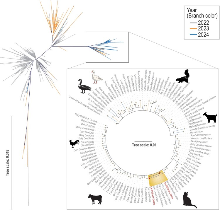
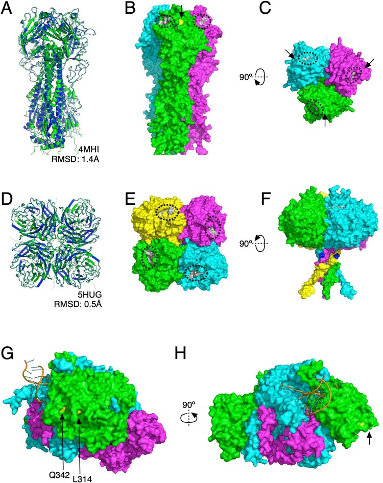
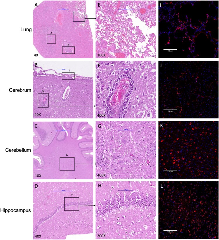
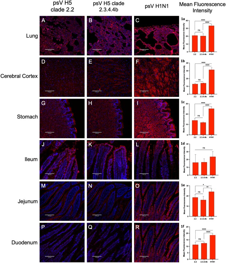
In April 2024, ten cats died in a rural South Dakota (SD) residence, showing respiratory and neurological symptoms. Necropsy and laboratory testing of two cats confirmed H5N1 clade 2.3.4.4b infection. The viral genome sequences are closely related to recent SD cattle H5N1 sequences. Cat H5N1 genomes had unique mutations, including T143A in haemagglutinin, known to affect infectivity and immune evasion, and two novel mutations in PA protein (F314L, L342Q) that may affect polymerase activity and virulence, suggesting potential virus adaptation. Dead cats showed systemic infection with lesions and viral antigens in multiple organs. Higher viral RNA and antigen in the brain indicated pronounced neurotropism. Lectin-histochemistry revealed widespread co-expression of sialic acid α-2,6 and α-2,3 receptors, suggesting cats could serve as mixing vessels for reassortment of avian and mammalian influenza viruses. No differences in clade 2.2 or 2.3.4.4b H5 pseudoviruses binding to cat lung/brain tissues indicated the neurotropism is unlikely mediated by receptor binding affinity.

Keywords: A(H5N1); Influenza A viruses; avian influenza; cat; clade 2.3.4.4b; influenza A virus evolution; neurotropism.


Conflict of interest statement

No potential conflict of interest was reported by the author(s).

Figures

Figure 1.
Figure 1.
Maximum likelihood-based haemagglutinin (HA) phylogenetic tree originating from 1443 sequences of HPAIV H5N1, clade 2.3.4.4b. The highlighted sub-branch of the unrooted tree contains the HPAIV H5N1 clade 2.3.4.4b sequences from two cats from South Dakota.
Figure 2.
Figure 2.
Exploration of mutational consequences on protein structure. (A) Side-view cartoon representation of the AlphaFold-predicted structure for HA trimer (green) superposed with a solved structure (PDB code: 4MHI; blue). (B) Surface representation of the predicted HA trimer structure, with subunits coloured in green, cyan, and magenta. The sialic acid binding pocket is circled and shaded grey near the top of the receptor-binding domain, and the nearby T143A mutation is in yellow with an arrow pointing to it. (C) The same but rotated 90 degrees to view from above the receptor-binding domain; all three sialic acid binding pockets and T143A positions are indicated. (D) Top-down view cartoon representation of the AlphaFold-predicted structure for NA tetramer (green) superposed with a solved structure (PDB code: 5HUG; blue). (E) The surface representation of the predicted NA tetramer structure, with subunits coloured green, cyan, magenta, and yellow. The sialic acid binding pocket is circled and shaded grey. (F) The same but rotated 90 degrees to view from the side; the location of the N71S mutation is indicated in blue. (G) Side-view surface representation of a viral polymerase complex previously solved by X-ray crystallography (8H69); the PA, PB1, and PB2 subunits are shaded green, cyan, and magenta, respectively. The locations of the mutations we observed in PA are shaded yellow. (H) The same but rotated 90 degrees to highlight the distance of observed mutations from the catalytic core of the polymerase complex.
Figure 3.
Figure 3.
Histopathological and immunohistochemical analysis of HPAIV H5N1 infected cat tissue sections. H&E-stained tissue sections of HPAIV H5N1 infected cat showed various histologic lesions. (A) Lung exhibited (1) interstitial pneumonia, (2) bronchiolitis, (3) bronchitis. (B) Cerebrum had (4) meningitis and (5) encephalitis whereas (D) hippocampus showed no lesions. The immunohistochemical (IHC) analysis (I-L) revealed the presence of IAV nucleoprotein in each of these organs. Brain tissues showed a higher nucleoprotein staining level than the lung tissue, with the cerebellum and hippocampus exhibiting extensive presence of nucleoprotein. Tissues were primarily stained using an anti-influenza A virus nucleoprotein antibody, followed by a secondary goat anti-mouse IgG H&L (Alexa Fluor® 647) antibody (red) and DAPI nuclear stain (blue). The H&E-stained images were acquired by Epredia Pannoramic MIDI II using Z stacking with extended focus, and the IHC images were acquired by ECHO revolve microscope.
Figure 4.
Figure 4.
Co-expression of SA α-2,3-Gal and SA α-2,6-Gal receptors in cat tissues. The composite fluorescent images reveal co-expression of SA α-2,3-Gal (red) and SA α-2,6-Gal (green) influenza receptors in the lung, cerebral cortex, stomach, ileum, jejunum, and duodenum of the cat. The lung and cerebral cortex exhibit co-expression of these receptors, whereas the gastrointestinal tissues display a higher expression of SA α-2,6-Gal (green) receptors, particularly on the luminal mucosal lining of the ileum, jejunum, and duodenum. Tissue sections were stained with FITC-labeled SNA (SA α-2,6-Gal) lectin, FITC-labeled MAA I (SA α-2,3-Gal), biotinylated MAA II (SA α-2,3-Gal), and DAPI nuclear stain (blue). Panels 1a–1f present the quantified mean fluorescence intensity for each corresponding tissue. Bars represent the mean ± standard deviation (SD) for each group (***p < 0.001, ****p < 0.0001). Mock-treated images are available in the supplementary data. Scale bar = 170 µm.
Figure 5.
Figure 5.
Widespread binding of pseudoviruses carrying H5 of clade 2.2 or clade 2.3.4.4b and human influenza H1 to cat tissues. The fluorescent images illustrate the extensive binding of H5N1 clade 2.2, 2.3.4.4b and human H1 pseudoviruses to cat tissues, including the (A,B,C) lung; (D,E,F) cerebral cortex; (G,H,I) stomach; (J,K,L) ileum; (M,N,O) jejunum; and (P,Q,R) duodenum. This binding pattern correlates with the relative abundance of SA α-2,3-Gal and SA α-2,6-Gal receptors in these tissues. The mucosal endothelial cells lining the alveoli and the luminal mucosal cells of the intestine exhibit a strong preference for virus binding. The tissue sections were primarily stained with mouse monoclonal anti-influenza virus H5 haemagglutinin (HA) protein (VN04-8), A/Vietnam/1203/2004 (H5N1) or anti-influenza A H1N1 haemagglutinin antibody followed by secondary goat anti-mouse IgG H&L (Alexa Fluor® 647) antibody (red) and DAPI nuclear stain (blue). Panel 1A-1F shows the quantified mean fluorescence intensity for each virus binding. Bars represent the mean ± standard deviation (SD) for each group (*p < 0.05, **p < 0.01, ****p < 0.0001). Mock-treated images are provided in the supplementary data. Scale bar = 170 µm.

References

    1. Gu M, Liu W, Cao Y, et al. . Novel reassortant highly pathogenic avian influenza (H5N5) viruses in domestic ducks, China. Emerg Infect Dis. 2011;17:1060–1063. doi:10.3201/eid/1706.101406 - DOI - PMC - PubMed
    1. Lewis NS, Banyard AC, Whittard E, et al. . Emergence and spread of novel H5N8, H5N5 and H5N1 clade 2.3.4.4 highly pathogenic avian influenza in 2020. Emerging Microbes Infect. 2021;10:148–151. doi:10.1080/22221751.2021.1872355 - DOI - PMC - PubMed
    1. Kandeil A, Patton C, Jones JC, et al. . Rapid evolution of A(H5N1) influenza viruses after intercontinental spread to North America. Nat Commun. 2023;14:3082. doi:10.1038/s41467-023-38415-7 - DOI - PMC - PubMed
    1. Burrough ER, Magstadt DR, Petersen B, et al. . Highly pathogenic avian influenza A(H5N1) Clade 2.3.4.4b virus infection in domestic dairy cattle and cats, United States, 2024. Emerg Infect Dis. 2024;30:1335–1343. doi:10.3201/eid3007.240508 - DOI - PMC - PubMed
    1. Blushke C. Influenza A (H5N1) virus detected in Wild Foxes (Vulpes vulpes) in Ontario. Healthy Wildlife, The blog of the Canadian Wildlife Health Cooperative, 2022.

MeSH terms

Substances