

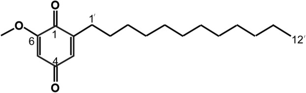
2-dodecyl-6-methoxycyclohexa-2,5-diene-1,4-dione mediates the effect of ROS-enhanced PI3K/Akt/mTOR pathway on autophagy in breast cancer
- PMID: 39648951
- PMCID: PMC11891764
- DOI: 10.1002/2211-5463.13940
2-dodecyl-6-methoxycyclohexa-2,5-diene-1,4-dione mediates the effect of ROS-enhanced PI3K/Akt/mTOR pathway on autophagy in breast cancer
Abstract
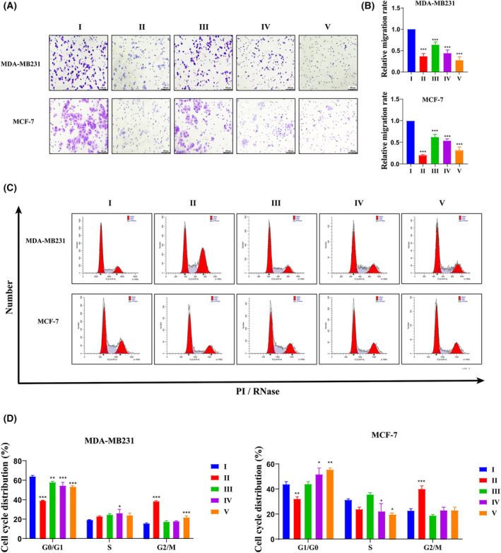
Several studies have suggested a potential antitumor effect of 2-dodecyl-6-methoxycyclohexa-2,5-diene-1,4-dione (DMDD). To further understand the mechanism of action of this compound, we investigated its effect on the phosphatidylinositol-3-kinase (PI3K)/serine-threonine kinase (Akt)/mammalian target of rapamycin (mTOR) signaling pathway. We show that DMDD application significantly inhibited the proliferation of breast cancer cell lines MDA-MB-231 and ER-α positive MCF-7. Furthermore, DMDD application resulted in increased intracellular reactive oxygen species (ROS) levels, apoptosis and autophagy, whereas it downregulated the expression of PI3K, Akt and mTOR mRNA and proteins, and increased the expression of LC3II/I and p62 proteins. In a mouse breast cancer xenograft model, DMDD inhibited tumor growth. Expression analyses suggest that ROS levels were higher in DMDD treated tumor tissues, whereas immunohistochemical analyses suggest that apoptotic cells were more prevalent in the DMDD treated group compared to the control group. Taken together, our results suggest that the molecular mechanism of action of DMDD may involve the enhancement of breast cancer autophagy through the PI3K/Akt/mTOR signaling pathway by mediating ROS expression.
Keywords: 2‐dodecyl‐6‐methoxycyclohexa‐2,5‐diene‐1,4‐dione; PI3K/Akt/mTOR signaling pathway; ROS; autophagy; breast cancer.
© 2024 The Author(s). FEBS Open Bio published by John Wiley & Sons Ltd on behalf of Federation of European Biochemical Societies.
Conflict of interest statement
The authors declare that they have no conflicts of interest.
Figures


References
-
- Siegel RL, Giaquinto AN and Jemal A (2024) Cancer statistics, 2024. CA Cancer J Clin 74, 12–49. - PubMed
-
- Bray F, Laversanne M, Sung H, Ferlay J, Siegel RL, Soerjomataram I and Jemal A (2024) Global cancer statistics 2022: GLOBOCAN estimates of incidence and mortality worldwide for 36 cancers in 185 countries. CA Cancer J Clin 74, 229–263. - PubMed
-
- Sung H, Ferlay J, Siegel R, Laversanne M, Soerjomataram I, Jemal A and Bray F (2021) Global cancer statistics 2020: GLOBOCAN estimates of incidence and mortality worldwide for 36 cancers in 185 countries. CA Cancer J Clin 71, 209–249. - PubMed
-
- Boon M and Akkari L (2024) Architecture sets the path: breast cancer subtypes differently shape the early brain metastatic niche. Cancer Cell 42, 1643–1645. - PubMed
MeSH terms
Substances
Grants and funding
- 2023KY0304/the Guangxi Zhuang Autonomous Region Education Department, the young and middle-aged teachers' basic scientific research ability improvement project
- ATYSP2023009/Autonomous Region Department of Science and Technology, ASEAN Outstanding Young Scientists Working in Guangxi China Project
- G202002014/Guangxi Medical High Level Bone Thousand Talents "139" Plan Training Personnel Washing Training objectives and responsibilities
- RCYJ202204/Guangxi University of Traditional Chinese Medicine Affiliated International Zhuang Medicine Hospital, talent introduction research start-up funding project
LinkOut - more resources
Full Text Sources
Medical
Miscellaneous
