[Preprint]. 2024 Nov 26:rs.3.rs-5486087.
doi: 10.21203/rs.3.rs-5486087/v1.

Distributed Temporal Coding of Visual Memory Categories in Human Hippocampal Neurons

Affiliations

Distributed Temporal Coding of Visual Memory Categories in Human Hippocampal Neurons

Xiwei She et al. Res Sq. .

Update in

Abstract

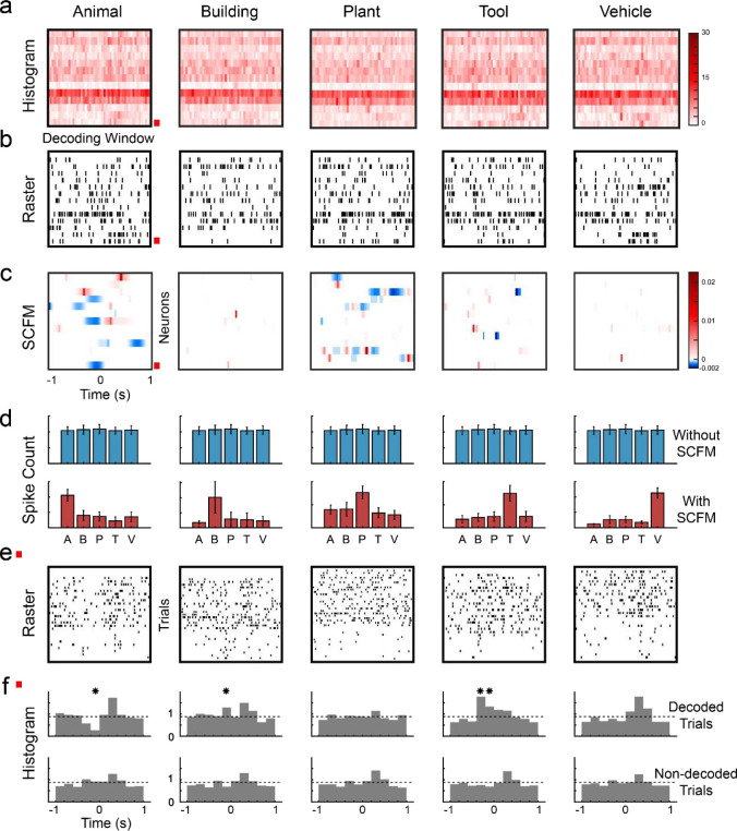
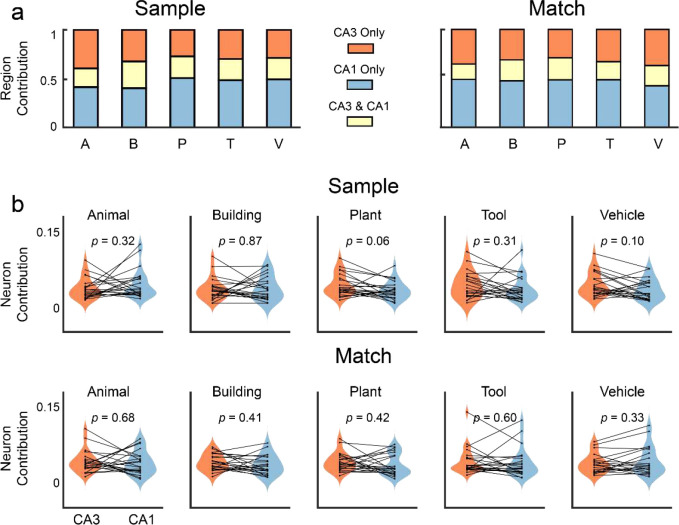
The hippocampus is crucial for forming new episodic memories. While the encoding of spatial and temporal information (where and when) in the hippocampus is well understood, the encoding of objects (what) remains less clear due to the high dimensions of object space. Rather than encoding each individual object separately, the hippocampus may instead encode categories of objects to reduce this dimensionality. In this study, we developed and applied a combined experimental-modeling approach to investigate how the hippocampus encodes visual memory categories in humans. We recorded spikes from hippocampal CA3 and CA1 neurons in 24 epilepsy patients performing a visual delayed match-to-sample (DMS) task involving five image categories. An ensemble multi-temporal-resolution classification model was employed to decode these visual memory categories from the hippocampal spiking activity with moderate numbers of trials. This model enables the identification of the spatio-temporal characteristics of hippocampal encoding through its interpretable representations. Using this model, we estimated the optimal temporal resolutions for decoding each visual memory category for each neuron in the ensemble. Results indicate that visual memory categories can be decoded from hippocampal spike patterns despite the short data length, supporting the presence of category-specific coding in the human hippocampus. We found that hippocampal neuron ensembles encode visual memory categories in a distributed manner, akin to a population code, while individual neurons use a temporal code. Additionally, CA3 and CA1 neurons exhibit similar and redundant information regarding visual memory categories, likely due to the strong and diffuse feedforward synaptic connections from the CA3 region to the CA1 region.

Keywords: Human hippocampus; memory category; memory decoding model; neurons; spatio-temporal code; spike.


Figures

Figure 1:
Figure 1:
Decoding visual memory categories from spatio-temporal patterns of spikes recorded in the human hippocampus using an ensemble multi-temporal-resolution classification model. This model provides interpretable model representations of spatio-temporal characteristics of spike patterns for encoding specific memory categories.
Figure 2:
Figure 2:
Categories of sample images can be decoded from spatio-temporal patterns of spikes recorded during Sample Response and Match Response events of a delayed DMS task in human subjects (n=24). a: DMS task paradigm. SP: Sample Presentation; SR: Sample Response; MP: Match Presentation; MR: Match Response. b: decoding cases and control cases in the modeling. c: decoding performance of the five memory categories. Color lines: MCCs of individual subjects; Black thick lines: average MCCs across all subjects; White shades: distributions of MCCs within categories.
Figure 3:
Figure 3:
Spatio-temporal distributions of category information in hippocampal spike patterns are revealed by the classification model. a: peri-event histogram of spike patterns of the five categories during the Sample Response events. b: raster plots showing a single trial of the spatio-temporal patterns of spikes during the Sample Response event. The five categories cannot be easily distinguished in either a or b. c: SCFMs of the five categories in the classification model. The red box marks the neuron shown in e and f. Based on the SCFMs, this neuron contributes to encoding the five categories. d: spike counts of each category with (top panel) and without (bottom panel) using SCFMs as masks. SCFMs reveal the spatio-temporal regions of the spike patterns that encode the category information. e: spike raster plots of trials within each category of the neuron marked in a, b, and c. f: peri-event histograms of trials of the decoded category (top panel) and other (non-decoded) categories (bottom panel) of this neuron. Dashed lines represent the baseline firing rates. Significant differences in firing rates between decoded categories and non-decoded categories exist in time intervals consistent with the SCFMs (bins marked with asterisks, p<0.05).
Figure 4:
Figure 4:
Sparseness of spatio-temporal encoding of visual memory categories during Sample Response and Match Response events of the DMS task. a: spatial sparseness of all neuron ensembles (n=24). b: temporal sparseness of all neurons (n=721). Bars: mean sparseness; error bars: standard deviation (STD) of sparseness. c: contribution of temporal resolutions to the encoding of visual memory categories. Colored dots (left y-axis): contribution of temporal resolutions to the encoding of categories in each subject. Colored lines (right y-axis): averaged contribution of temporal resolutions across all subjects (n=24). Black line (right y-axis): averaged contribution of temporal resolutions across all five categories. Left: Sample Response; Right: Match Response.
Figure 5:
Figure 5:
Contribution of hippocampal CA3 and CA1 to the encoding of visual memory categories during Sample Response and Match Response events of the DMS task. a: Contributions of the CA3 and CA1 regions. Red: unique contribution of the CA3 region; Yellow: redundant contribution shared by CA3 and CA1 regions; Blue: unique contribution of the CA1 region. b: averaged contribution of hippocampal CA3 and CA1 neurons to the encoding. Each dot represents one subject (n=24). There is no significant difference between CA3 and CA1 neurons during both Sample Response and Match Response (paired t-test).
Figure 6.
Figure 6.
Human experimental paradigm. a: 3T MRI showing pre-implantation hippocampal structures and post-implantation electrode locations in one subject. Inset: zoomed-in view of the probe in the hippocampus. b: Layout of the micro-macro probe containing 6 macro-electrodes and 10 micro-electrodes. Six and four micro-electrodes were implanted in the CA3 and CA1 regions, respectively. c: Sample Images of the five memory categories used in the DMS task.

References

    1. Izquierdo I. & Medina J. H. Memory formation: the sequence of biochemical events in the hippocampus and its connection to activity in other brain structures. Neurobiol. Learn. Mem. 68, 285–316 (1997). - PubMed
    1. Deadwyler S. A. & Hampson R. E. Ensemble activity and behavior: What’s the code? Science (1995) doi: 10.1126/science.270.5240.1316. - DOI - PubMed
    1. Burgess N., Maguire E. A. & O’Keefe J. The human hippocampus and spatial and episodic memory. Neuron 35, 625–641 (2002). - PubMed
    1. Scoville W. B. & Milner B. Loss of recent memory after bilateral hippocampal lesions. J. Neurol. Neurosurg. Psychiatry 20, 11 (1957). - PMC - PubMed
    1. Bigler E. D. et al. Traumatic brain injury and memory: the role of hippocampal atrophy. Neuropsychology 10, 333 (1996).

Publication types

LinkOut - more resources