[Preprint]. 2024 Nov 29:2024.11.28.24318055.
doi: 10.1101/2024.11.28.24318055.

Blood methylation biomarkers are associated with diabetic kidney disease progression in type 1 diabetes

Affiliations

Blood methylation biomarkers are associated with diabetic kidney disease progression in type 1 diabetes

Anna Syreeni et al. medRxiv. .

Abstract

Background: DNA methylation differences are associated with kidney function and diabetic kidney disease (DKD), but prospective studies are scarce. Therefore, we aimed to study DNA methylation in a prospective setting in the Finnish Diabetic Nephropathy Study type 1 diabetes (T1D) cohort.

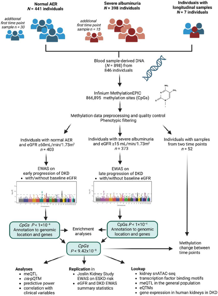
Methods: We analysed baseline blood sample-derived DNA methylation (Illumina's EPIC array) of 403 individuals with normal albumin excretion rate (early progression group) and 373 individuals with severe albuminuria (late progression group) and followed-up their DKD progression defined as decrease in eGFR to <60 mL/min/1.73m2 (early DKD progression group; median follow-up 13.1 years) or end-stage kidney disease (ESKD) (late DKD progression group; median follow-up 8.4 years). We conducted two epigenome-wide association studies (EWASs) on DKD progression and sought methylation quantitative trait loci (meQTLs) for the lead CpGs to estimate genetic contribution.

Results: Altogether, 14 methylation sites were associated with DKD progression (P<9.4×10⁻⁸). Methylation at cg01730944 near CDKN1C and at other CpGs associated with early DKD progression were not correlated with baseline eGFR, whereas late progression CpGs were strongly associated. Importantly, 13 of 14 CpGs could be linked to a gene showing differential expression in DKD or chronic kidney disease. Higher methylation at the lead CpG cg17944885, a frequent finding in eGFR EWASs, was associated with ESKD risk (HR [95% CI] = 2.15 [1.79, 2.58]). Additionally, we replicated meQTLs for cg17944885 and identified ten novel meQTL variants for other CpGs. Furthermore, survival models including the significant CpG sites showed increased predictive performance on top of clinical risk factors.

Conclusions: Our EWAS on early DKD progression identified a podocyte-specific CDKN1C locus. EWAS on late progression proposed novel CpGs for ESKD risk and confirmed previously known sites for kidney function. Since DNA methylation signals could improve disease course prediction, a combination of blood-derived methylation sites could serve as a potential prognostic biomarker.


Figures

Figure 1.
Figure 1.. Study setting.
Abbreviations: AER=albumin excretion rate; cis-pQTM = cis protein quantitative trait methylation; DKD=diabetic kidney disease; EWAS=epigenome-wide association study; eGFR=estimated glomerular filtration rate; eQTMs=expression quantitative trait methylations; meQTL=methylation quantitative trait locus; snATAC-seq=single-nucleus transposase-accessible chromatin with sequencing. Created in BioRender. Syreeni, A. (2024) https://BioRender.com/.
Figure 2
Figure 2. Manhattan plots show the results of EWASs on DKD progression.
A) Results from the EWAS on early DKD progression, B) early DKD progression EWAS additionally adjusted for the baseline eGFR, and C) results from the EWAS on late DKD progression (to ESKD). X-axis shows the chromosomal position and y-axis shows the −log10 of the association P-value. Methylation sites reaching epigenome-wide significance (P<9.4×10⁻⁸, green line) are annotated into the plot.
Figure 3.
Figure 3.. Methylation site cg01730944 is located close to CDKN1C.
A) Density plot of early DKD progression cohort (n=403) baseline methylation beta values of cg01730944 shows lower methylation in individuals with progressing DKD during follow-up [eGFR decline <60 mL/min/1.73 m2 (in orange)] compared to individuals who do not progress (light blue) B) Kaplan–Meier plot compares individuals in the lowest and highest tertile for cg01730944 methylation and shows the proportion of individuals progressing to eGFR<60 mL/min/1.73 m2 during follow-up. C) Open chromatin peaks in kidney cell types; human kidney single-nucleus transposase-accessible chromatin data (Version 2) on 57,229 cells accessed in Susztaklab Kidney Biobank. Figure is adapted from https://susztaklab.com/Human_snATAC/, and cg01730944 position is incorporated. D) Kidney single-cell expression data of 23,980 nuclei shows that CDKN1C is mainly expressed in podocytes. Adapted from Humphrey’s Lab browser at http://humphreyslab.com E) In vivo expression of CDKN1C in human glomerular cells shows lower expression (fold-change=−4.95, P=4.9×10⁻⁵ in diabetic kidney disease (group 2, n=9) compared to individuals without DKD (group 1, n=13). Figure adapted from Nephroseq v.5 database at https://www.nephroseq.org/. Abbreviations: PT-S1–PT-S3=proximal tubule segments 1–3; LOH=loop of Henle; DCT=distal convoluted tubule; PC=principal cells of collecting duct; IC=intercalated cells, Endo=endothelia; Podo=podocytes; Immune=immune cells; lymph=lymphocytes; MES=mesenchyme, PEC=parietal epithelial cell; PCT=proximal convoluted tubule; DCT/CT=distal convoluted tubule/connecting tubule; CD-PC=collecting duct - principal cell; CD-ICA=collecting duct - intercalated cells A; CD-ICB=collecting duct - intercalated cells B; Leuk=leukocytes
Figure 4.
Figure 4.. Predictive power of the lead CpGs.
The diamonds show the concordance (C-index) and its 95% confidence intervals of three Cox proportional-hazards models applied for the early (n=393 with non-missing variables) and late DKD progression (n=363 with non-missing variables) cohorts. P-values denote the significance of the increase in concordance index compared to the previous model; The significant P-values (P<0.05) are marked in the figure. The first model, “Clinical variables” (orange color), included baseline triglyceride concentration, central obesity, and current smoking status for the early DKD progression analysis, and triglyceride concentration, HbA1c, and systolic blood pressure for the late DKD progression analysis. Additionally, the model included six white blood cell proportions, technical PCs 1–3, mean methylation M value from invariable sites, age, and sex. The second model (red color) included additionally baseline eGFR. The third model included methylation M values for four (early DKD progression-associated: cg25013571, cg05831784, cg06334496, and cg01730944) or nine (late DKD progression-associated: cg06536988, cg03262246, cg11115840, cg21871803, cg14999724, cg10579797, cg04166335, cg12272104, and cg17944885) methylation sites.
Figure 5.
Figure 5.. Links between methylation and gene expression of trans-meQTL locus on chromosome 16.
According to Huan et al, SNV rs17611866 correlates (in trans) with methylation levels of 45 CpGs, of which eGFR-associated methylation sites cg17944885 (chr19p13.2 locus, in multiple EWASs) and cg06158227 are shown in the figure. CpG cg17944885 has also a close SNV rs4804653 that is associated with its methylation levels in the general population (GoDMC) data. We replicated both the cis- and trans-methylation quantitative trait loci in our diabetes cohort. Abbreviations: cis-eQTL=cis expression quantitative trait locus (SNV that affects gene expression); cis-meQTL=cis methylation quantitative trait locus; trans-meQTL=trans methylation quantitative trait locus (SNV that associates with CpG site methylation); cis-eQTM=cis-expression quantitative trait methylation (methylation site that associates with gene expression). Created in BioRender. Syreeni, A. (2024) https://BioRender.com/.

References

    1. Jansson Sigfrids F, Groop PH, Harjutsalo V. Incidence rate patterns, cumulative incidence, and time trends for moderate and severe albuminuria in individuals diagnosed with type 1 diabetes aged 0-14 years: a population-based retrospective cohort study. Lancet Diabetes Endocrinol. 2022;10(7):489–498 doi:10.1016/S2213-8587(22)00099-7 - DOI - PubMed
    1. Salem RM, Todd JN, Sandholm N, Cole JB, Chen WM, Andrews D, et al. Genome-Wide Association Study of Diabetic Kidney Disease Highlights Biology Involved in Glomerular Basement Membrane Collagen. J Am Soc Nephrol. 2019;30(10):2000–2016 doi: 10.1681/ASN.2019030218 - DOI - PMC - PubMed
    1. Sandholm N, Cole JB, Nair V, Sheng X, Liu H, Ahlqvist E, et al. Genome-wide meta-analysis and omics integration identifies novel genes associated with diabetic kidney disease. Diabetologia. 2022;65(9):1495–1509 doi:10.1007/s00125-022-05735-0 - DOI - PMC - PubMed
    1. Sandholm N, Dahlström EH, Groop PH. Genetic and epigenetic background of diabetic kidney disease. Front Endocrinol. 2023;14:1163001 doi:10.3389/fendo.2023.1163001 - DOI - PMC - PubMed
    1. Bell CG, Teschendorff AE, Rakyan VK, Maxwell AP, Beck S, Savage DA. Genome-wide DNA methylation analysis for diabetic nephropathy in type 1 diabetes mellitus. BMC Med Genomics. 2010;3:33 doi:10.1186/1755-8794-3-33 - DOI - PMC - PubMed

Publication types