Case Reports
. 2024 Nov 22:14:1465676.
doi: 10.3389/fonc.2024.1465676. eCollection 2024.

Primary intracranial malignant melanoma in an adolescent girl with NRAS and TP53 mutations: case report and literature review

Affiliations
Case Reports

Primary intracranial malignant melanoma in an adolescent girl with NRAS and TP53 mutations: case report and literature review

Xinyu Liu et al. Front Oncol. .

Abstract

Primary intracranial malignant melanoma(PIMM) is often difficult to treat in patients without a history of skin melanoma or extensive melanin deposition. Due to the rarity of the disease, the current accepted treatment is surgical resection, but the prognosis is still poor. We report a case of PIMM in an adolescent girl with epilepsy as the only symptom and atypical imaging findings. PIMM was confirmed by further pathological and clinical examination. We summarize previous cases to discuss the clinical manifestations, imaging, pathological and genetic characteristics of the disease, aiming to improve the clinician's understanding of the disease. This case underscores the PIMM as a differential diagnosis and prompt surgical treatment for adolescents with epileptic seizures accompanied by intracranial space-occupying lesions, even in the absence of extensive skin blackening.

Keywords: NRAS; adolescent; case report; nevus; pediatric; primary malignant melanoma; seizures.


Conflict of interest statement

The authors declare that the research was conducted in the absence of any commercial or financial relationships that could be construed as a potential conflict of interest.

Figures

Figure 1
Figure 1
Several small speckled black pigmentations were seen in the left axilla (A). The pathological findings of skin nevus suggested benign intradermal nevus (B). Intraoperatively seen (C).The immunohistochemical staining: HMB45 + (D), S100 + (E).The pathological findings of skin nevus suggested benign intradermal nevus (F).
Figure 2
Figure 2
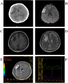
CT shows high-density opacities such as round-like images (A),DWI high signal (B),T1WI high-signal (C), T2WI low-signal shadow (D), the Cr peak was lower, the Cho peak was higher, the NAA peak was not shown, and the Cho/Cr was higher (E, F).
Figure 3
Figure 3
Postoperative CT (A) and MRI (B).
Figure 4
Figure 4
Screening process for past cases.

Similar articles

Cited by

References

    1. Louis DN, Perry A, Reifenberger G, von Deimling A, Figarella-Branger D, Cavenee WK, et al. . The 2016 world health organization classification of tumors of the central nervous system: a summary. Acta Neuropathol. (2016) 131:803–20. doi: 10.1007/s00401-016-1545-1 - DOI - PubMed
    1. Byun J, Park ES, Hong SH, Cho YH, Kim Y-H, Kim CJ, et al. . Clinical outcomes of primary intracranial Malignant melanoma and metastatic intracranial Malignant melanoma. Clin Neurol Neurosurg. (2018) 164:32–8. doi: 10.1016/j.clineuro.2017.11.012 - DOI - PubMed
    1. Farabi B, Akay BN, Goldust M, Wollina U, Atak MF, Rao B. Congenital melanocytic naevi: An up-to-date overview. Australas J Dermatol. (2021) 62:e178–91. doi: 10.1111/ajd.13535 - DOI - PubMed
    1. Mjaatvedt CH, Kern CB, Norris RA, Fairey S, Cave CL. Normal distribution of melanocytes in the mouse heart. Anat Rec A Discovery Mol Cell Evol Biol. (2005) 285:748–57. doi: 10.1002/ar.a.20210 - DOI - PubMed
    1. Uehara S, Izumi Y, Kubo Y, Wang CC, Mineta K, Ikeo K, et al. . Specific expression of Gsta4 in mouse cochlear melanocytes: a novel role for hearing and melanocyte differentiation. Pigment Cell Melanoma Res. (2009) 22:111–9. doi: 10.1111/j.1755-148X.2008.00513.x - DOI - PubMed

Publication types

LinkOut - more resources