Review
. 2024;7(6):825-856.
doi: 10.1007/s42242-024-00316-z. Epub 2024 Oct 23.

Biofabrication and biomanufacturing in Ireland and the UK

Affiliations
Review

Biofabrication and biomanufacturing in Ireland and the UK

Jack F Murphy et al. Biodes Manuf. 2024.

Abstract

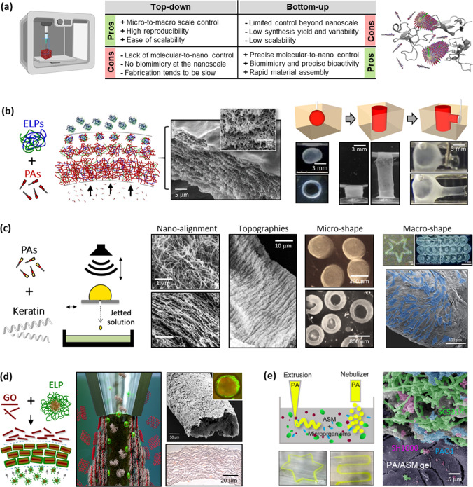
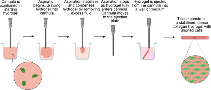
As we navigate the transition from the Fourth to the Fifth Industrial Revolution, the emerging fields of biomanufacturing and biofabrication are transforming life sciences and healthcare. These sectors are benefiting from a synergy of synthetic and engineering biology, sustainable manufacturing, and integrated design principles. Advanced techniques such as 3D bioprinting, tissue engineering, directed assembly, and self-assembly are instrumental in creating biomimetic scaffolds, tissues, organoids, medical devices, and biohybrid systems. The field of biofabrication in the United Kingdom and Ireland is emerging as a pivotal force in bioscience and healthcare, propelled by cutting-edge research and development. Concentrating on the production of biologically functional products for use in drug delivery, in vitro models, and tissue engineering, research institutions across these regions are dedicated to innovating healthcare solutions that adhere to ethical standards while prioritising sustainability, affordability, and healthcare system benefits.

随着我们从第四次工业革命向第五次工业革命的过渡,生物制造领域的兴起正在改变生命科学和医疗保健。3D生物打印、组织工程、定向组装和自组装等先进技术在创建仿生支架、组织类器官、医疗设备和生物系统方面发挥着重要作用。英国和爱尔兰的生物制造领域成为生物科学和医疗保健的关键力量,得益于尖端的研究和发展。这些地区的研究专注于药物输送、体外模型, 类器官,和组织工程的生物功能产品,致力于创新符合伦理标准的医疗保健解决方案,同时支持环保和有效的医疗保健系统。.

Keywords: Biohybrid; Biomaterials; Bioprinting; Drug delivery; Sustainability; Tissue engineering.


Conflict of interest statement

Conflict of interestYYSH is an associate editor for Bio-Design and Manufacturing and was not involved in the editorial review or the decision to publish this article. The authors declare that they have no conflict of interest.

Figures

Fig. 1
Fig. 1
A non-exhaustive regional breakdown of biofabrication and biomanufacturing groups from around the UK and Ireland as of March 2024
Fig. 2
Fig. 2
Next generation of implants: design of selective glyco-functionalised polymers will enable attachment of sugars to the end-groups of the polymer back bone to induce immunomodulation facilitating a desirable host response
Fig. 3
Fig. 3
A potential biofabrication pathway for engineering complex osteochondral grafts. a Microtissues are biofabricated in a high-throughput fashion using micromoulding, whereby single cells self-organise into spherical cellular aggregates. Phenotypically distinct microtissues can be generated by using different cell types or by exposing aggregated stem cells to tissue-specific culture conditions. b Once formed, microtissue laden bioinks are deposited via bioprinting to create spatially organised constructs. c Following bioprinting, adjacent microtissues fuse to form a single macrotissue which is matured in vitro to create tissue rudiments for articular cartilage and subchondral bone. Once implanted, the precursor tissues continue to mature, mirroring the native postnatal osteochondral developmental program, resulting in the formation of a functional osteochondral unit and regeneration of the joint surface
Fig. 4
Fig. 4
Applications of polymers and polymer nanocomposites in biomedical engineering and healthcare. a Diagram showing some important applications of polymers and polymer nanocomposites: b microspheres for drug delivery and injection therapy (reproduced from Ref. [73], Copyright 2015, with permission from The Royal Society of Chemistry), c bilayered porous scaffolds for tissue engineering (reproduced from Ref. [75], Copyright 2021, with permission from the authors, licensed under CC BY 4.0), d hydrogel microfibres (reproduced from Ref. [77], Copyright 2017, with permission from the American Chemical Society), e a pH-sensitive polymer (reproduced from Ref. [76], Copyright 2018, with permission from The Royal Society of Chemistry), f a self-healing polymer nanocomposite as a flexible electronic device (reproduced from Ref. [85], Copyright 2016, with permission from the American Chemical Society), and g a strain sensor for monitoring joint movements (reproduced from Ref. [86], Copyright 2017, with permission from the American Chemical Society)
Fig. 5
Fig. 5
Supramolecular biofabrication. a Advantages and disadvantages of merging top-down (e.g., biofabrication) and bottom-up (e.g., supramolecular self-assembly) strategies. Reproduced from Ref. [106], Copyright 2010, with permission from Elsevier. b Through diffusion–reaction mechanisms, the supramolecular assembly of peptide amphiphiles (PAs) and elastin-like proteins (ELPs) gives rise to hierarchical-organised membranes and allows liquid-in-liquid fabrication of dynamic tubular structures. Reproduced from Ref. [99] (Copyright 2015, with permission from Macmillan Publishers Limited) and Ref. [107] (Copyright 2023, with permission from the authors, licensed under CC BY 4.0). c Drop-on-demand printing, with its involved hydrodynamic forces, is exploited to create PA-keratin composites with dictated fibre nano-alignment, surface topography, and macroscopic geometries. Reproduced from Ref. [102], Copyright 2018, with permission from WILEY-VCH Verlag GmbH & Co. KGaA, Weinheim. d Graphene oxide (GO) flakes co-assembled with ELPs are harnessed to grow and bioprint vascular-like perfusable tubes. Reproduced from Ref. [103] (Copyright 2020, with permission from the authors, licensed under CC BY 4.0) and Ref. [104] (Copyright 2021, with permission from the authors, licensed under CC BY 4.0). e Co-assembly of PAs and artificial sputum enables the creation of printable 3D models of bacterial infection. Reproduced from Ref. [105], Copyright 2023, with permission from the authors, licensed under CC BY 4.0
Fig. 6
Fig. 6
Fabrication of corneal hydrogels. a Poly-epsilon-lysine forms the basis of the hydrogels (pεK). b pεK is methacrylated by reacting m pεK with methacrylic anhydride and triethylamine at pH 7 and 20 °C for 12 h to produce pεKMA. Adapted from Ref. [108] (Copyright 2014, with permission from the authors, licensed under CC BY 4.0) and Ref. [109] (Copyright 2018, with permission from the authors, licensed under CC BY 3.0). c A co-polymer is formed from pεK and poly(ethylene glycol) diacrylate (PEGDA). d The mechanical properties are improved for co-polymer hydrogels compared to pεK and PEGDA alone. Cell attachment is affected by the formulation of the hydrogel as demonstrated using a human corneal endothelial cell line (HCEC12 cells). HCEC12 cells on e 100% pεK hydrogel, f 90% pεK/10% PEGDA, g 80% pεK/20% PEGDA, and h 50% pεK/50% PEGDA, after 7 d in culture (ZO-1 red, Phalloidin green, DAPI blue). The mechanical properties of the hydrogel are optimal for handling and delivery. i The hydrogel being delivered to the rabbit eye using an intraocular lens injector device and j after easy unfolding and centration in the anterior chamber. k The excellent positioning and attachment of the graft to the posterior rabbit cornea can be seen using optical coherence tomography (red indicating cornea and green graft). DAPI: 4',6-diamidino-2-phenylindole
Fig. 7
Fig. 7
Pericytes embedded in hydrogel support the barrier function of brain microvascular endothelial cells (BMECs). OX1-19 induced pluripotent stem cells (iPSCs) were differentiated into BMECs and pericytes as described [112, 113]. PureCol hydrogel was layered on the surface of a Transwell insert with a coating of Matrigel on the upper surface. a BMECs were either cultured as a monoculture on top of the hydrogel, or mixed with pericytes on top of the hydrogel, or the pericytes were encapsulated within the hydrogel with the BMECs layered on top. Transendothelial electrical resistance (TEER) measurements were taken daily for five days and plotted as b unit area resistance (UAR) on respective days, c area under the curve (AUC) of b, and d peak UAR of b. TEER measurements were taken using an EVOM2 voltmeter and STX3 electrodes (World Precision Instruments, UK). Peak UAR is the highest recording of TEER of the whole measured time period. AUC and peak UAR were analysed using one-way analysis of variance (ANOVA) with Tukey’s multiple comparisons test. Data are shown as mean ± SEM. *P 0.05, **P 0.01. SEM: standard error of the mean
Fig. 8
Fig. 8
Biomanufacturing using microdroplet and protein nanosheet technologies. a Schematic representation of engineered protein nanosheets. Reproduced from Ref. [131], Copyright 2024, with permission from the authors, licensed under CC BY. b Protein nanosheet assembly can be orchestrated in microfluidic platforms to control the size of microdroplets. c Examples of protein nanosheet designs. Adapted from Ref. [124], Copyright 2023, with permission from the authors, licensed under CC BY 4.0. d Transmission electron microscopy images of protein nanosheets (albumin-based). e Changes in interfacial shear mechanics taking place upon assembly of poly(L-lysine) nanosheets at a liquid–liquid interface. Reproduced from Ref. [122], Copyright 2022, with permission from the authors, licensed under CC BY 4.0. f Brightfield microscopy image of HEK293 cells growing on a bioemulsion. g Colony of mesenchymal stem cells growing on a microdroplet (blue, nuclei; red, F-actin; green, vinculin). Reproduced from Ref. [123], Copyright 2023, with permission from the authors, licensed under CC BY 4.0. h Schematic representation of a microdroplet-based bone marrow microenvironment. Reproduced from Ref. [133], Copyright 2023, with permission from the authors, licensed under CC BY 4.0
Fig. 9
Fig. 9
Intestinal tissue engineering—progress to date and future directions. a Schematic depicting intestinal structure and function, the predominant engineering strategies to date and proposed future directions. The intestine, a hollow tubular organ, has an inner mucosa, consisting of epithelium with supporting mesenchyme, a sub-mucosa containing vasculature and lymphatics, surrounded by circular and longitudinal muscle layers regulated by an enteric nervous system. It has various key functions including digestion and absorption, transit via peristalsis and the maintenance of a barrier against luminal micro-organisms. b, c Predominant intestinal engineering strategies to date include full-thickness tissue-engineered small intestine (TESI) from human intestinal organoids (HIOs) and mucosal grafts engineered from human intestinal stem cell (ISC) organoids. d Future directions include 3D biofabrication and culture techniques to increase engineered graft size and mucosal repurposing to generate a segment of small intestinalised colon. Figure created using BioRender.com
Fig. 10
Fig. 10
Flow diagram of the gel aspiration-ejection (GAE) method for biofabrication of engineered tissue constructs. Figure created using BioRender.com
Fig. 11
Fig. 11
Multiscale biofabrication techniques. a Bio-assembling macro-scale, lumised airway tubes (reproduced from Ref. [158], Copyright 2021, with permission from the authors, licensed under CC BY). b Suspended piezoelectric nanofibre networks for acoustic sensing. c 3D printing of soft biomaterials. d 3D printing suspended fibres for cell culture (reproduced from Ref. [163], Copyright 2019, with permission from the American Chemical Society). e Microfluidic cell culture with microvessels (reproduced from Ref. [154], Copyright 2021, with permission from the authors, licensed under CC BY 3.0). f Cell ‘morphing’ on extracellular matrix analogues (reproduced from Ref. [164], Copyright 2014, with permission from the authors, licensed under CC BY)
Fig. 12
Fig. 12
Engineered nanotopographies as minimally invasive nanotools for cargo delivery, biosensing, and in vivo cell reprogramming. a Schematic representation of the diverse applications of high-aspect-ratio nanostructured surfaces which leverage intimate contact between the substrate material and cell membrane (reproduced from Ref. [165], Copyright 2020, with permission from WILEY–VCH Verlag GmbH & Co. KGaA, Weinheim). b Nanoneedles have been employed to deliver specific cargo which elucidated uptake mechanisms: b1 focused-ion-beam scanning electron microscope (SEM) imaging demonstrated cell membrane interaction with nanostructures with the formation of two classes of endocytic vesicles clathrin pits (orange arrows) and caveolae (green arrows) (scale bars: 100 nm); b2 the organisation of vesicle structures on nanoneedles (red) and non-nanoneedle locations (blue) was achieved using 3D reconstruction. Reproduced from Ref. [166], Copyright 2019, with permission from the authors, licensed under CC BY. c Nanoneedles have been used to detect cancerous cells (OE 33) based on cytosolic levels of cathepsin B (CTSB): c1 representative images of a single z-plane through the cytosol of CTSB − ve (HET-1A) and + ve (OE 33) cells where yellow fluorescence arises from CTSB mediated cleavage of a fluorescent probe on the nanopatterned substrate and blue is the nuclei (scale bars: 25 µm); c2 quantification of the area-normalised cytosolic fluorescence signal for OE 33 (yellow) and HET-1A (blue) when interfaced with nanoneedle sensors for various times (x-axis). Reproduced from Ref. [171], Copyright 2015, with permission from the authors, licensed under CC BY. d Nanoneedles have enabled the in vivo delivery of a growth-factor-encoding plasmid. d1 Bright-field (top) and confocal (bottom) microscopy showing vasculature within muscles of untreated (control) and human vascular endothelial growth factor (hVEGF)-165 treated (direct-injection and nanoinjection). Fluorescent signal is from systemically injected fluorescein isothiocyanate (FITC)-dextran (scale bars: 100 µm for bright-field and 50 µm for confocal). Vessel quantification is demonstrated by d2 the fraction of fluorescent signal and d3 the number of nodes in the vasculature per mm2. Reproduced from Ref. [167], Copyright 2015, with permission from Macmillan Publishers Limited
Fig. 13
Fig. 13
Harnessing nanoclay chemistry for regenerative medicine. a The synthetic clay Laponite consists of 25-nm diameter, 1-nm thick disks that possess a permanent negative surface charge and a pH dependent rim charge (positive below pH 9) (reproduced from Ref. [214], Copyright 2013, with permission from WILEY–VCH Verlag GmbH & Co. KGaA, Weinheim). b These properties generate a surprisingly rich array of possibilities for regenerative medicine. b1 Aqueous solutions of nanoclay self-assemble into stiff gels upon contact with blood to form bioactive environments able to attract the invasion of stem cells ​(reproduced from Ref. [215], Copyright 2018, with permission from WILEY–VCH Verlag GmbH & Co. KGaA, Weinheim)​. b2 The addition of nanoclay within hydrogel composites offers improved rheological properties as well as cell viability (reproduced from Ref. [201], Copyright 2019, with permission from IOP Publishing Ltd).​ b3 Due to their affinity for proteins such as growth factors, nanoclay gels can sustain localised concentrations to promote new tissue formation in the body at reduced effective doses, and controlling diffusion reaction processes allows for high-resolution control over stable protein concentration gradients for direct tissue regeneration​ (the left part was reproduced from Ref. [202], Copyright 2020, with permission from the authors, licensed under CC BY 4.0; the right part was reproduced from Ref. [209], Copyright 2023, with permission from the authors, licensed under CC BY)
Fig. 14
Fig. 14
Acoustic cell patterning techniques and applications. a Fluorescence microscopy of acoustically patterned myoblasts, demonstrating the flexibility and rapid dynamic control of ultrasound patterning. Scale bars: 200 µm. Reproduced from Ref. [35], Copyright 2019, with permission from the authors, licensed under CC BY. b Histology showing the deposition of aligned collagen fibres by acoustically-patterned primary bovine articular chondrocytes in agarose: b1 staining for sulfated glycosaminoglycan using safranin O, b2 immunostaining for type II collagen, b3, b4 polarized light microscopy following picrosirius red staining. Scale bars: 100 µm for b1, b2, 50 µm for b3, and 5 µm for b4. Reproduced from Ref. [220], Copyright 2022, with permission from the authors, licensed under CC BY. c Distinct regions of angiogenic sprouting (c1, c2) and cellular network formation (c3, c4) in differentially acoustically patterned regions of RFP-HUVEC-laden bioprinted constructs. Scale bars: 100 µm. Reproduced from Ref. [221], Copyright 2023, with permission from IOP Publishing Ltd. d Acoustic holographic patterning of HCT-116 cells within a collagen hydrogel. Scale bar: 5 mm; inset scale bar: 500 µm. Reproduced from Ref. [223], Copyright 2019, with permission from the authors, licensed under CC BY. e In vivo acoustic patterning of collagen-suspended GFP-HUVECs (reproduced from Ref. [226], Copyright 2023, with permission from the authors, licensed under CC BY 4.0). HUVEC: human umbilical vein endothelial cell; GFP: green fluorescent protein; RFP: red flourescent protein
Fig. 15
Fig. 15
Biofabrication research in Shu’s lab: a 3D-bioprinted droplet array of human embryo stem cells (reproduced from Ref. [229], Copyright 2013, with permission from IOP Publishing Ltd); b 3D-printed robust, freestanding alginate hydrogel blood vessel-like structure using secondary (Ca2+) and tertiary (Ba2+) crosslinking steps (scale bar: 20 p coin) (reproduced from Ref. [231], Copyright 2015, with permission from the authors, licensed under CC BY 3.0); c 3D-printed polypeptide-DNA hydrogel with blue dye added for visualisation that is robust enough to be picked up by tweezers (reproduced from Ref. [232], Copyright 2015, with permission from Wiley–VCH Verlag GmbH & Co. KGaA, Weinheim); d 3D-reconstructed confocal laser scanning microscope Z-stack of P. aeruginosa biofilm in a 4-mm thick hydrogel construct following 14 days of maturation (scale bar: 100 µm) (reproduced from Ref. [233], Copyright 2019, with permission from the authors, licensed under CC BY 3.0); e multilayered alginate hydrogel tubular structure produced via a micro-dip coating method (scale bar: 20 p coin) (reproduced from Ref. [234], Copyright 2017, with permission from the authors, licensed under CC BY); f fluorescence image of a collagen-organoid tube for transplantable bile duct applications (scale bar: 100 µm) (reproduced from Ref. [236], Copyright 2017, with permission from Nature America, Inc., part of Springer Nature); g confocal image of multilayer prelabelled red, green, and blue high density human embryonic kidney (HEK) cells in a 1% (0.01 g/mL) alginate tube positioned via the rotational internal flow engineering (RIFLE) method (scale bar: 100 µm) (reproduced from Ref. [237], Copyright 2023, with permission from the authors, licensed under CC BY 4.0); h live/dead fluorescent imaging of murine adipose derived stem cells within a micromachined, electrospun polylactic-co-glycolic acid (PLGA) fibre mesh (scale bar: 500 µm) (reproduced from Ref. [238], Copyright 2023, with permission from the authors, licensed under CC BY 4.0)

References

    1. Xu M, David JM, Kim SH (2018) The Fourth Industrial Revolution: opportunities and challenges. Int J Financ Res 9(2):90–95. 10.5430/ijfr.v9n2p90
    1. Office for National Statistics (2024) National Life Tables—Life Expectancy in the UK: 2020 to 2022. Statistical Bulletin. https://www.ons.gov.uk/peoplepopulationandcommunity/birthsdeathsandmarri...
    1. Bosurgi R (2019) Climate crisis: healthcare is a major contributor, global report finds. BMJ 336:l5560. 10.1136/bmj.l5560 - PubMed
    1. Global Carbon Project (2019) Supplemental Data of Global Carbon Project 2019. Global Carbon Project. 10.18160/GCP-2019
    1. Taylor K, Alvarez LR (2019) An estimate of the number of animals used for scientific purposes worldwide in 2015. Altern Lab Anim 47(5–6):196–213. 10.1177/0261192919899853 - PubMed

LinkOut - more resources