. 2024 Nov 22:12:1491962.
doi: 10.3389/fbioe.2024.1491962. eCollection 2024.

Engineered myeloid precursors differentiate into osteoclasts and resorb heterotopic ossification in mice

Affiliations

Engineered myeloid precursors differentiate into osteoclasts and resorb heterotopic ossification in mice

Cameron Rementer et al. Front Bioeng Biotechnol. .

Abstract

Introduction: Heterotopic ossification (HO) occurs following orthopedic trauma, spinal cord injuries, brain trauma and limb amputations. Once symptomatic, HO causes pain, limited mobility and decreased quality of life. Current treatments are limited and have significant complications with high recurrence rates, underscoring the need for improved therapeutic interventions. Osteoclasts (OCs) are physiological bone resorptive cells that secrete enzymes and protons to degrade bone.

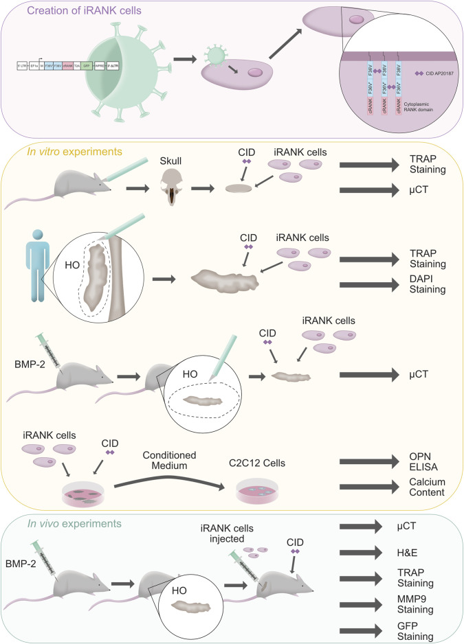
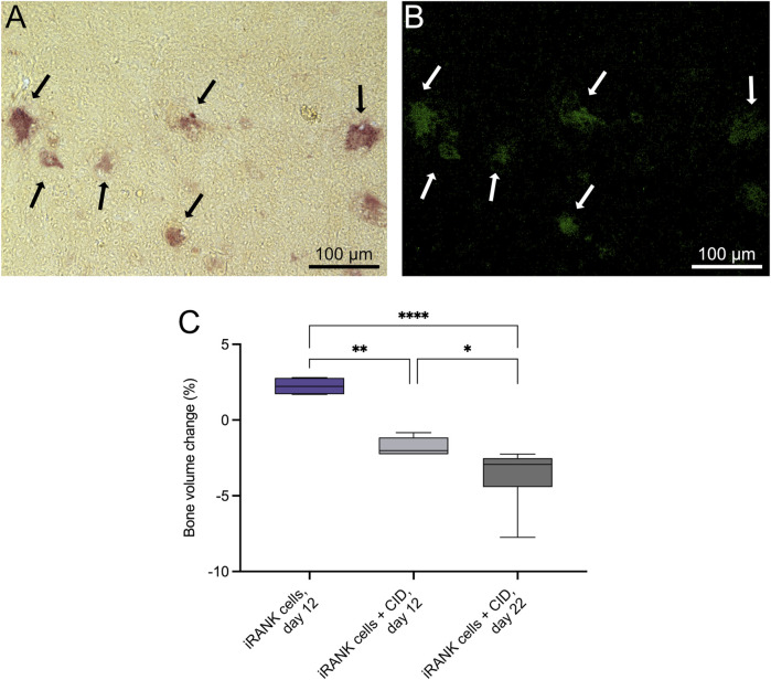
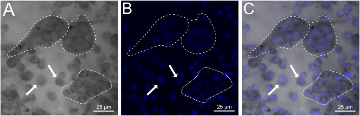
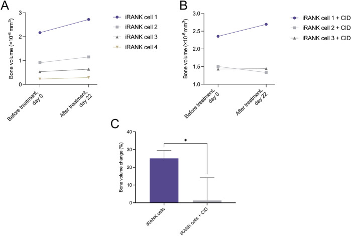
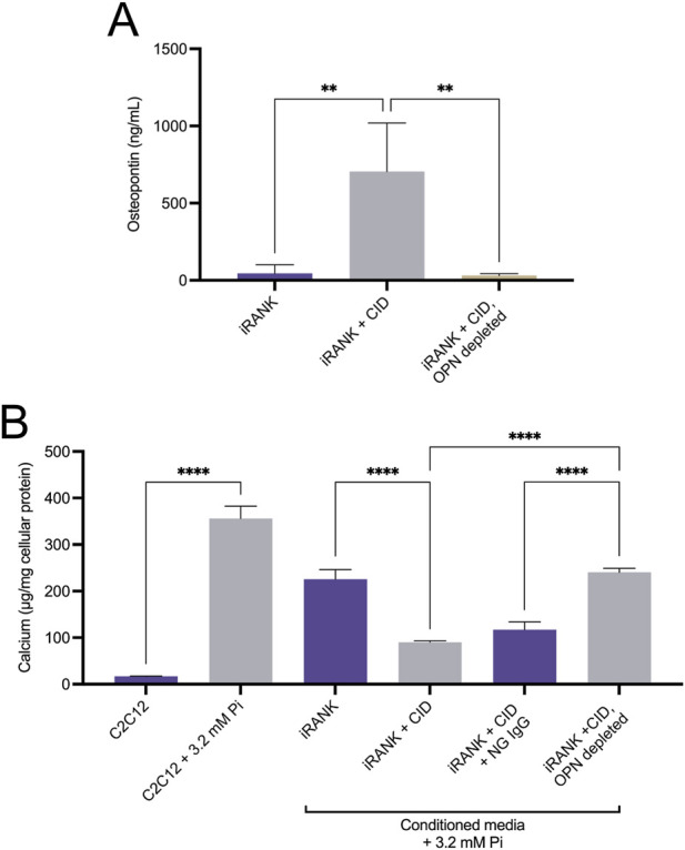
Methods: In this study, we describe the use of genetically engineered OCs as a novel cell therapy approach to treat HO. Inducible, engineered myeloid precursors (iRANK cells) treated with a chemical inducer of dimerization (CID) differentiated into TRAP+ multinucleated OCs and resorbed mineralized tissues in vitro.

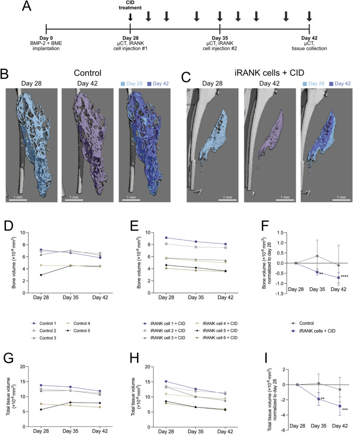
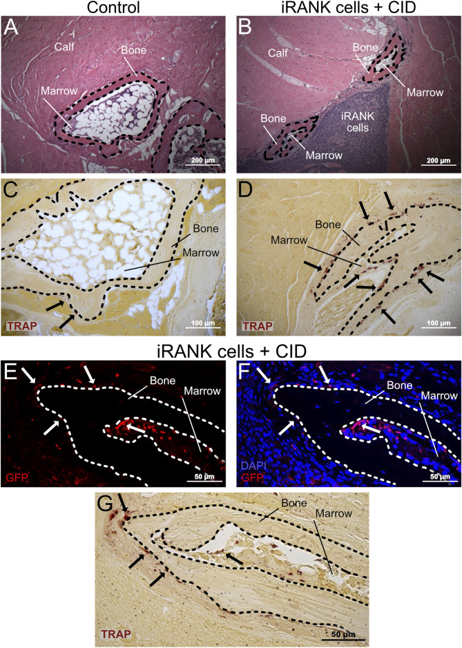
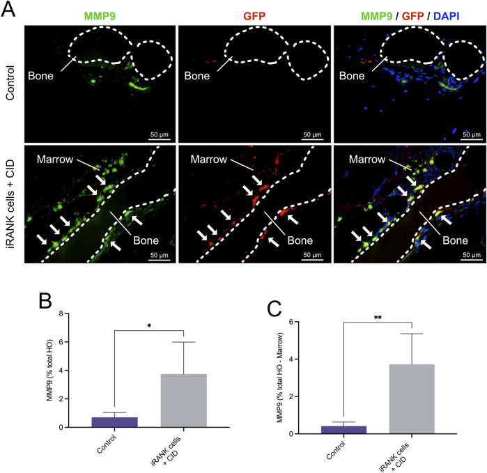
Results: In vivo, BMP-2-induced murine HO lesions were significantly regressed following treatment using iRANK cells with concomitant systemic administration of CID. Moreover, many OCs were TRAP+, MMP9+, and GFP+, indicating that they differentiated from delivered iRANK cells.

Discussion: In summary, these data con rm the ability of engineered myeloid precursors to differentiate into OCs and resorb HO in vivo paving the way for OC delivery as a promising approach for HO treatment.

Keywords: RANK; bone; chemical inducer of dimerization; engineered osteoclasts; heterotopic ossification; osteoclasts; resorption.


Conflict of interest statement

The authors declare that the research was conducted in the absence of any commercial or financial relationships that could be construed as a potential conflict of interest. The author(s) declared that they were an editorial board member of Frontiers, at the time of submission. This had no impact on the peer review process and the final decision.

Figures

FIGURE 1
FIGURE 1
Outline of the study. A schematic summary of the creation of engineered osteoclasts (iRANK cells) is shown with a purple background. These cells were tested within in vitro (yellow background) and in vivo (green background) experiments. Illustration drawn by Hannah Blümke using Affinity Designer 2.1.1.
FIGURE 2
FIGURE 2
iRANK cells induced with a chemical inducer of dimerization resorb calvarial bone in vitro. Following osteoclast differentiation of iRANK cells using a chemical inducer of dimerization, osteoclasts (A) stain for TRAP (black arrows), and (B) also appear GFP+ (white arrows) since engineered iRANK cells contain a GFP domain within the iRANK construct. (C) µCT analysis of in vitro treated murine calvarial disks showed a significant decrease in the percentage of bone volume change compared to disks treated with iRANK cells that had not been induced using the chemical inducer of dimerization. This decrease could be enhanced by reseeding and continued induction of cells using the chemical inducer of dimerization, as assessed by Šídák’s multiple comparison in conjunction with ANOVA (n = 4–7 donors, *p < 0.05, **p < 0.01, ****p < 0.0001). Scale bar: A, B = 100 µm.
FIGURE 3
FIGURE 3
iRANK cells adhere to and differentiate into osteoclasts on human heterotopic ossifications. (A) Brightfield confocal imaging showing diffuse as well as punctate cytoplasmic TRAP staining in mononucleated pre-osteoclasts (white arrows) and multinucleated osteoclasts (dashed white line). Preosteoclasts in the process of fusion are outlined by a dotted white line. (B) DAPI staining confirms multinucleation of osteoclasts (dashed white line), which are also depicted additionally in a (C) merged image. Scale bar = 25 µm.
FIGURE 4
FIGURE 4
Murine heterotopic ossification nodules significantly decreased in the percentage of bone volume change in response to treatment with activated iRANK cells. (A) Overall, an increasing trend in bone volume can be observed for nodules seeded with iRANK cells, but not activated using the chemical inducer of dimerization, probably due to passive mineralization. (B) When differentiating and activating iRANK cells using the chemical inducer of dimerization two out of three cell groups show a decreasing trend in relation to bone volume. (C) Data expressed as the percentage of bone volume change demonstrate a statistically significant reduction, as assed by Student’s t-test (n = 3–4, *p < 0.05).
FIGURE 5
FIGURE 5
Functional, phosphorylated OPN is highly secreted by engineered osteoclasts. (A) iRANK cells were cultured in the absence or presence of 50 nM chemical inducer of dimerization to induce osteoclast differentiation. ELISA was used to detect osteopontin (OPN) levels in the culture media. Untreated iRANK cells secreted significantly less OPN than iRANK cells induced with the chemical inducer of dimerization. OPN was successfully depleted from conditioned media using an OP-199 antibody, as assessed by Tukey’s multiple comparison in conjunction with ANOVA (n = 3–4, **p < 0.01). (B) Conditioned media was transferred every day to C2C12 cells in the presence of 3.2 mM inorganic phosphate for 5 days. Conditioned media from iRANK cells induced with the chemical inducer of dimerization inhibited C2C12 calcification. Removal of OPN with OP-199 antibody eliminated the inhibitory effect of the conditioned media from iRANK cells induced with the chemical inducer of dimerization. Normal goat IgG (NG) was used as an isotype control for OP-199 antibody, as assessed by Tukey’s multiple comparison in conjunction with ANOVA (n = 3, ****p < 0.0001).
FIGURE 6
FIGURE 6
Resorption of HO formations by engineered osteoclasts in vivo. (A) Experimental timeline showing cell delivery, chemical inducer of dimerization injection, and µCT time schedules following induction of HO formations in calf muscle with BMP-2 and a basement membrane extract (BME). (B) µCT images show HO formations, which appear stable with little change in overall morphology over time in the control group, while (C) a significant decrease in the size of the HO formation could be seen following treatment using iRANK cells and the chemical inducer of dimerization. (D) A stable trend in bone volume can be seen in the control group, while (E) animals injected with iRANK cells and the chemical inducer of dimerization depict a decreasing trend. (F) When normalized to day 28, a statistically significant decrease can be seen in the treatment group as assessed by Tukey’s multiple comparison in conjunction with ANOVA (n = 5–6, **p < 0.01, ****p < 0.0001). (G) The total tissue volume was also stable with only slight decreases in the control group while (H) a larger decrease in total tissue volume could be seen in the treatment group. (I) When normalized to day 28, a significant decrease could also be observed regarding total tissue volume in the treatment group, as assessed by Tukey’s multiple comparison in conjunction with ANOVA (n = 5–6, **p < 0.01, ***p < 0.001).
FIGURE 7
FIGURE 7
Large numbers of engineered OCs were observed on HO formations in animals treated with iRANK cells and the chemical inducer of dimerization. H&E staining shows HO formations exhibiting a ring-like structures with a mineralized surface (B = Bone) surrounding a marrow-like cavity (M = Marrow) in the (A) control group as well as the (B) treatment group. (C) A greater number of TRAP+ osteoclasts (black arrowheads) were observed lining the HO formations in the treatment group compared to the (D) control group. (E) Immunofluorescence staining for GFP (in red) indicates that many of the (F) multinucleated cells (nuclei in blue) lining the HO formations were derived from locally delivered iRANK cells in the iRANK and chemical inducer of dimerization treatment group. (G) These GFP+ multinucleated cells were also TRAP+ as shown in an adjacent tissue section. Dashed lines outline bone tissue. Scale bar: (A, B) = 200 μm, (C, D) = 100 μm, (E–G) = 50 µm.
FIGURE 8
FIGURE 8
Immunofluorescence staining of MMP9+ osteoclasts in HO formations. (A) Double immunofluorescence staining showing numerous MMP9+GFP+ multinucleated OCs lining bone surfaces (B = Bone) with some MMP9+GFP+ mononuclear cells in the marrow spaces (M = Marrow) in the iRANK treatment group, while only a few MMP9+GFP endogenous osteoclasts were found in the control group. (B) MMP9+ cells were quantified as percentage of signal intensity per total HO area. (C) Results remained statistically significant after subtracting the marrow-like area in which predominantly MMP9+ mononuclear cells were found, as assessed by Student’s t-test (n = 3, *p < 0.05, **p < 0.01).

References

    1. Agarwal S., Loder S., Cholok D., Li J., Breuler C., Drake J., et al. (2017). Surgical excision of heterotopic ossification leads to Re-emergence of mesenchymal stem cell populations responsible for recurrence. Stem Cells Transl. Med. 6, 799–806. 10.5966/SCTM.2015-0365 - DOI - PMC - PubMed
    1. Ausk B. J., Gross T. S., Bain S. D. (2015). Botulinum toxin-induced muscle paralysis inhibits heterotopic bone formation. Clin. Orthop. Relat. Res. 473, 2825–2830. 10.1007/S11999-015-4271-4 - DOI - PMC - PubMed
    1. Blümke A., Ijeoma E., Simon J., Wellington R., Purwaningrum M., Doulatov S., et al. (2023). Comparison of osteoclast differentiation protocols from human induced pluripotent stem cells of different tissue origins. Stem Cell Res. Ther. 14, 319. 10.1186/S13287-023-03547-6 - DOI - PMC - PubMed
    1. Blümke A., Simon J., Leber E., Scatena M., Giachelli C. M. (2024). Differentiation and characterization of osteoclasts from human induced pluripotent stem cells. JoVE J. Vis. Exp. 2024, e66527. 10.3791/66527 - DOI - PMC - PubMed
    1. Bossche L. V., Vanderstraeten G. (2005). Heterotopic ossification: a review. J. Rehabil. Med. 37, 129–136. 10.1080/16501970510027628 - DOI - PubMed

LinkOut - more resources