Case Reports
. 2024 Nov 29;39(1):669.
doi: 10.4102/sajid.v39i1.669. eCollection 2024.

Brain abscesses in an immunocompromised patient with a soft tissue mass

Affiliations
Case Reports

Brain abscesses in an immunocompromised patient with a soft tissue mass

Thokozani Mwase et al. S Afr J Infect Dis. .

Abstract

Nocardiosis is a rare opportunistic infection and may be misdiagnosed as tuberculosis in the immunocompromised patient. This case report highlights the importance of doing tissue cultures in immunocompromised individuals to correctly identify Nocardia spp. and initiate appropriate treatment timeously.

Contribution: This case report describes a typical case of disseminated nocardiosis with brain abscesses in an immunocompromised patient who would have typically been treated as disseminated tuberculosis.

Keywords: CNS; HIV; Nocardia; brain abscess; immunocompromised; nocardiosis.


Conflict of interest statement

The authors declare that they have no financial or personal relationships that may have inappropriately influenced them in writing this article.

Figures
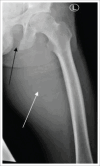
FIGURE 1
FIGURE 1
Left frontal thigh radiograph. Left thigh prominent soft tissue swelling (white arrow) associated with erosion of the left inferior pubic ramus (black arrow).
FIGURE 2
FIGURE 2
Frontal chest radiograph: left upper lung airspace opacity with internal cavitation (black arrow) and left axillary soft tissue swelling (white arrow).
FIGURE 3
FIGURE 3
Frontal radiograph of the left shoulder: Left axillary soft tissue prominence (white arrow). Left upper lobe air space disease, with early internal cavitation (black arrow).
FIGURE 4
FIGURE 4
(a) Left distal forearm lateral soft tissue swelling (white arrow). No bone erosion. Small wrist and forearm radio-densities are unchanged and likely related to prior trauma. (b) Frontal projection of the left lower limb demonstrated lateral soft tissue swelling (white arrow). No fibular erosion.
FIGURE 5
FIGURE 5
(a) Axial post-contrast computed tomography (CT) brain demonstrates vasogenic oedema (white arrow) and rim-enhancing lesions in the left parietal lobe (black arrows). (b) Sagittal post-contrast brain CT demonstrated loculated rim-enhancing lesions (black arrow), of which some are bilobed (white arrow).

References

    1. Trevisan V. I Generi E Le Specie Delle Batteriacee Milan: Zanaboni & Gabuzzi. Reprod Int Bull Bacteriol Nomencl Taxon. 1889;2:13–44.
    1. Fatahi-Bafghi M. Nocardiosis from 1888 to 2017. Microb Pathog. 2018;114:369–384. 10.1016/j.micpath.2017.11.012 - DOI - PubMed
    1. Meena DS, Kumar D, Bohra GK, Midha N, Garg MK. Clinical characteristics and treatment outcome of central nervous system nocardiosis: A systematic review of reported cases. Med Princ Pract. 2022;31(4):333–341. 10.1159/000525509 - DOI - PMC - PubMed
    1. Lerner PI. Nocardiosis. Clin Infect Dis. 1996;22(6):891–903. 10.1093/clinids/22.6.891 - DOI - PubMed
    1. Conville PS, Brown-Elliott BA, Smith T, Zelazny AM. The complexities of Nocardia taxonomy and identification. J Clin Microbiol. 2018;56(1):10–128. 10.1128/jcm.01419-17 - DOI - PMC - PubMed

Publication types

LinkOut - more resources