. 2024 Nov 22:15:1487726.
doi: 10.3389/fimmu.2024.1487726. eCollection 2024.

G. vaginalis increases HSV-2 infection by decreasing vaginal barrier integrity and increasing inflammation in vivo

Affiliations

G. vaginalis increases HSV-2 infection by decreasing vaginal barrier integrity and increasing inflammation in vivo

Nuzhat Rahman et al. Front Immunol. .

Abstract

Introduction: Clinically, a dysbiotic vaginal microbiota (VMB) colonized with anaerobic species such as Gardnerella vaginalis has been linked to increased susceptibility to viral sexually transmitted infections (STIs) such as Herpes Simplex Virus Type 2 (HSV-2). The mechanism is poorly understood due to the lack of small animal models.

Methods: Mice were inoculated with 107 CFU of the eubiotic bacteria Lactobacillus crispatus, the dysbiotic bacteria G. vaginalis, or PBS as a negative control every 48 h for ten days. On day ten, mice were inoculated with 105 PFU WT HSV-2 333 and survival, pathology, and viral titers were assessed. To elucidate changes in the vaginal microenvironment following bacterial inoculations, vaginal tissue and washes were collected following ten days of inoculations. To assess barrier integrity, tissue was fixed and stained for the barrier protein Desmoglein-1 (DSG-1). To evaluate the immune microenvironment, tissue was processed for flow cytometry to examine tissue-resident T cells and cytokine production by T cells. Vaginal washes were used for multiplex cytokine/chemokine analysis.

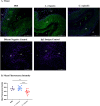
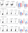
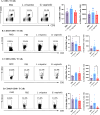
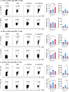
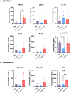
Results: G. vaginalis inoculated mice infected with HSV-2 had significantly decreased survival rates, increased pathology, and higher viral titers than PBS and L. crispatus inoculated mice. The vaginal epithelium of G. vaginalis inoculated mice showed decreased DSG-1 staining compared to other groups, indicating compromised barrier function. Decreased total numbers of CD4+ and CD8+ T cells expressing activated mucosal immune markers CD44, CD69, and CD103 were observed in the vaginal tract of G. vaginalis inoculated mice. They also showed increased proportions of T cells expressing inflammatory cytokines TNF-α and IFN-γ, while L. crispatus inoculated mice had increased proportions and absolute counts of T cells expressing the regulatory cytokine IL-10. In the multiplex assay, vaginal washes from G. vaginalis mice had increased inflammatory cytokines and chemokines compared to L. crispatus and PBS groups.

Discussion: These results suggest G. vaginalis inoculation may be increasing HSV-2 infection by disrupting the epithelial barrier, decreasing protective immune responses and increasing tissue inflammation in the vaginal tract.

Keywords: Lactobacillus; bacterial vaginosis; barrier integrity; female reproductive health; herpes simplex virus; inflammation; mouse models; vaginal microbiota (VMB).


Conflict of interest statement

The authors declare that the research was conducted in the absence of any commercial or financial relationships that could be construed as a potential conflict of interest.

Figures

Figure 1
Figure 1
L. crispatus and G. vaginalis were consistently colonized when inoculated every 48 h. Female mice were inoculated twice 48 h or 72 h apart with 107 CFU L. crispatus, G. vaginalis, or PBS as a negative control. Data are from n = 6 per group from one experiment representative of 3 independent experiments with similar results. Vaginal washes were collected post-inoculation for up to 48 h or 72 h after the second inoculation and assessed using quantitative plating assays. Bacterial colonies of the inoculated species types were counted in PBS inoculated 48 h apart (A), PBS inoculated 72 h apart (B), L. crispatus inoculated 48 h apart (C), L. crispatus inoculated 72 h apart (D), G. vaginalis inoculated 48 h apart (E), and G. vaginalis inoculated 72 hours apart (F) groups. Different coloured points denote different mice. The data was analyzed using a two-way ANOVA with Tukey’s multiple comparisons, but no significance was found.
Figure 2
Figure 2
G. vaginalis inoculated mice showed decreased survival rates, increased pathology, and higher viral titers when infected with HSV-2 compared to L. crispatus and PBS-inoculated mice. Mice were administered 107 CFU L. crispatus, G. vaginalis, or PBS as a no-exogenous bacteria-negative control every 48 h for 10 days. 24 h after the most recent inoculation, mice in diestrus were infected with 105 PFU wildtype HSV-2 333. Data in panels (A, B) include cumulative data from three independent experiments (n=18 in the PBS group, n=17 in the L. crispatus group, and n=19 in the G. vaginalis group). Mice were euthanized when they reached a pathology score of 4 or 5, and survival was graphed in panel (A). Vaginal washes were collected up to 7 days post-infection and viral titers were determined with a Vero plaque assay (B). Vaginal pathology was recorded up to 7 days post-infection. Individual mouse data from one independent experiment representative of the three experiments was plotted for pathology (C) and viral titers (D) (n=7 per group, except L. crispatus n=6). Different coloured points denote different mice in panels (C, D). Survival data was analyzed using a Log-rank (Mantel-Cox) test (**p<0.01). Cumulative viral titers were analyzed with a two-way ANOVA with Tukey’s multiple comparisons (****p<0.0001).
Figure 3
Figure 3
G. vaginalis inoculated mice had decreased Desmoglein-1 expression in the vaginal epithelium compared to L. crispatus and PBS groups. Mice were administered 107 CFU L. crispatus, G. vaginalis, or PBS as a no-exogenous bacteria-negative control every 48 h for 10 days. Vaginal tissue was collected from the mice and fixed in methacarn. The tissue was paraffin-embedded, deparaffinized, and stained for Desmoglein-1 (green) with a DAPI counterstain (blue). Images were taken using a confocal microscope at 20X magnification (A). The lumen (L), epithelium (EP), and lamina propria (LP) are indicated on all images. Mean fluorescence intensity was evaluated using three representative images per sample with ImageJ software (B). Data is representative of two independent experiments (n=13 per group, except PBS n=7). The data was analyzed using a one-way ANOVA with Tukey’s multiple comparisons (**p<0.01 and ****p<0.0001).
Figure 4
Figure 4
G. vaginalis inoculated mice had decreased activated and mucosal CD4+ T cells in the vaginal tract compared to L. crispatus inoculated mice. Female mice were intravaginally inoculated with 107 CFU L. crispatus, G. vaginalis, or PBS as a no-exogenous bacteria-negative control every 48 h for 10 days. On day 10 of the experiment, the vaginal tissue (VAG) was collected, processed, and stimulated for 16 h. Cells were stained for Live/Dead staining, CD3, CD4, CD44, CD69, and CD103 and ran on the Cytoflex flow cytometer and analyzed using FloJo software. The percent population and absolute count of CD4+ T cells are shown in panel (A). The percent population and absolute number of CD44+CD4+ T cells (B), CD69+CD4+ T cells (C), and CD103+CD4+ T cells (D) are depicted as well. Data are from n=5 per group, from one experiment representing three independent experiments. Data was analysed using a one-way ANOVA with Tukey’s multiple comparisons (*p<0.05).
Figure 5
Figure 5
G. vaginalis inoculated mice had decreased activated and mucosal CD8+ T cells in the vaginal tract compared to L. crispatus inoculated mice. Female mice were intravaginally inoculated with 107 CFU L. crispatus, G. vaginalis, or PBS as a no-exogenous bacteria-negative control every 48 h for 10 days. On day 10 of the experiment, the vaginal tissue (VAG) was collected, processed, and stimulated for 16 h. Cells were stained for Live/Dead staining, CD3, CD8, CD44, CD69, and CD103, and ran on the Cytoflex flow cytometer and analyzed using FloJo software. The percent population and absolute number of CD8+ T cells are shown in panel (A). The percent population and absolute number of CD44+CD8+ T cells (B), CD69+CD8+ T cells (C), and CD103+CD8+ T cells (D) are depicted as well. Data are from n=5 per group, from one experiment representing three independent experiments. Data was analyzed using a one-way ANOVA with Tukey’s multiple comparisons (*p<0.05).
Figure 6
Figure 6
G. vaginalis inoculated mice had increased T cells expressing inflammatory cytokines and L. crispatus treated mice had increased T cells expressing regulatory cytokines in the vaginal tract. Female mice were intravaginally inoculated with 107 CFU L. crispatus, G. vaginalis, or PBS as a no-exogenous bacteria-negative control every 48 h for 10 days. On day 10 of the experiment, the vaginal tissue (VAG) was collected, processed, and stimulated for 16 h. Cells were stained for Live/Dead staining, CD3, CD4, CD8, TNF-α, IFN-γ, and IL-10, and ran on the Cytoflex flow cytometer and analyzed using FloJo software. CD4+ and CD8+ T cells expressing TNF-α (A), IFN-γ (B), and IL-10 (C) are depicted. Data are from n=5 per group, from one experiment representing three independent experiments. Data was analyzed using a one-way ANOVA with Tukey’s multiple comparisons (*p<0.05 and **p<0.01).
Figure 7
Figure 7
G. vaginalis inoculated mice had increased inflammatory cytokine and chemokine protein levels in the vaginal tract. Female mice were intravaginally inoculated with 107 CFU L. crispatus, G. vaginalis, or PBS as a no-exogenous bacteria-negative control every 48 h for 10 days. On day 10 of the experiment, vaginal washes were collected and sent to Eve Technologies to perform a 31-Plex mouse cytokine/chemokine assay. All significant cytokine (A) and chemokine (B) data are shown above. Data are from n=5 per group, from one independent experiment. Data was analyzed using a one-way ANOVA with Tukey’s multiple comparisons (*p<0.05, **p<0.01, and ***p<0.001).

References

    1. Peric A, Weiss J, Vulliemoz N, Baud D, Stojanov M. Bacterial colonization of the female upper genital tract. Int J Mol Sci. (2019) 20:3405. doi: 10.3390/ijms20143405 - DOI - PMC - PubMed
    1. Ravel J, Gajer P, Abdo Z, Schneider GM, Koenig SSK, McCulle SL, et al. . Vaginal microbiome of reproductive-age women. Proc Natl Acad Sci. (2011) 108:4680–7. doi: 10.1073/pnas.1002611107 - DOI - PMC - PubMed
    1. Chee WJY, Chew SY, Than LTL. Vaginal microbiota and the potential of lactobacillus derivatives in maintaining vaginal health. Microbial Cell Factories. (2020) 19:203. doi: 10.1186/s12934-020-01464-4 - DOI - PMC - PubMed
    1. Ghartey JP, Smith BC, Chen Z, Buckley N, Lo Y, Ratner AJ, et al. . Lactobacillus crispatus dominant vaginal microbiome is associated with inhibitory activity of female genital tract secretions against escherichia coli. PloS One. (2014) 9:e96659. doi: 10.1371/journal.pone.0096659 - DOI - PMC - PubMed
    1. Chen X, Lu Y, Chen T, Li R. The female vaginal microbiome in health and bacterial vaginosis. Front Cell Infect Microbiol. (2021) 11. doi: 10.3389/fcimb.2021.631972 - DOI - PMC - PubMed