. 2024 Nov 8;27(12):111350.
doi: 10.1016/j.isci.2024.111350. eCollection 2024 Dec 20.

Epigenetic control of myogenic identity of human muscle stem cells in Duchenne muscular dystrophy

Affiliations

Epigenetic control of myogenic identity of human muscle stem cells in Duchenne muscular dystrophy

Jimmy Massenet et al. iScience. .

Abstract

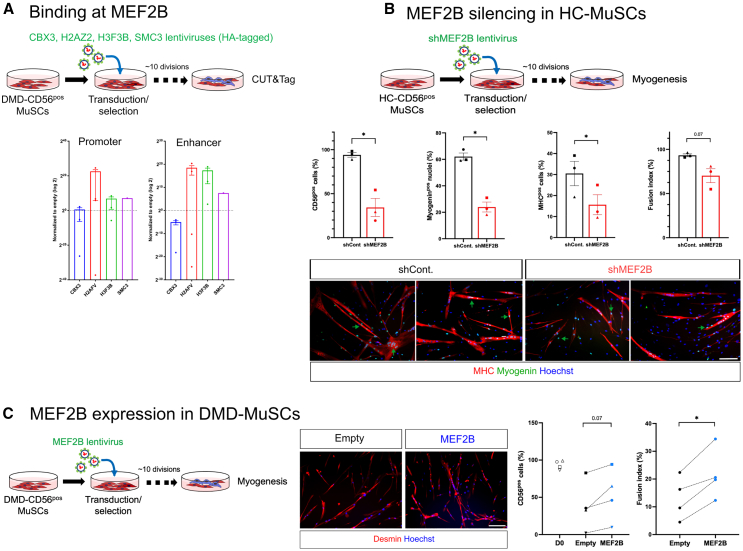
In Duchenne muscular dystrophy (DMD), muscle stem cells' (MuSCs) regenerative capacities are overwhelmed leading to fibrosis. Whether MuSCs have intrinsic defects or are disrupted by their environment is unclear. We investigated cell behavior and gene expression of MuSCs from DMD or healthy human muscles. Proliferation, differentiation, and fusion were unaltered in DMD-MuSCs, but with time, they lost their myogenic identity twice as fast as healthy MuSCs. The rapid drift toward a fibroblast-like cell identity was observed at the clonal level, and resulted from altered expression of epigenetic enzymes. Re-expression of CBX3, SMC3, H2AFV, and H3F3B prevented the MuSC identity drift. Among epigenetic changes, a closing of chromatin at the transcription factor MEF2B locus caused downregulation of its expression and loss of the myogenic fate. Re-expression of MEF2B in DMD-MuSCs restored their myogenic fate. MEF2B is key in the maintenance of myogenic identity in human MuSCs, which is altered in DMD.

Keywords: Epigenetics; Integrative aspects of cell biology; Stem cells research.


Conflict of interest statement

Authors declare no competing interests.

Figures

None
Graphical abstract
Figure 1
Figure 1
In vitro behavior of DMD-MuSCs (A–C) CD56pos MuSCs isolated from healthy control (HC) and Duchenne (DMD) muscles were analyzed for their capacity to implement in vitro myogenesis. (A) Proliferation was assessed in growth medium (GM) as the number of EdUpos cells (red). (B) Differentiation was quantified after 5 days in differentiation medium (DM) as the number of myogeninpos cells (green) among desminpos cells (red). (C) Fusion index was quantified in differentiated cells grown at high density, as the number of nuclei in desmin expressing myotubes (red) related to the total number of nuclei. Hoechst labels nuclei (blue). Bar: 100 μm. (D) Expression of CD56 was evaluated by flow cytometry during the culture of initially pure CD56pos HC- and DMD-MuSCs in growth medium. (E) Calculation of the loss of CD56 per cell division from (D). (F) Experimental procedure of the clonal culture of Myf5-transduced CD56pos MuSCs. (G) Immunostaining of clones for CD56 (red), GFP(Myf5) (green) and TCF7L2 (cyan). Hoechst labels nuclei (blue). Red arrowheads show myogenic CD56posMyf5pos cells, blue arrowheads show fibrogenic TCF7L2pos cells and yellow arrowheads show cells harboring both myogenic and fibrogenic markers. Bar: 50 μm. (H) Quantification of cells according to the immunostaining shown in (G). Each shape symbol represents one clone. Results are means ± SEM of 3–9 samples in (A–E) and of 6 clones issued from 3 HC donors and of 5 clones issued from 3 DMD donors in (H). ns: non significant, ∗p < 0.05 using unpaired (A–E) or paired (H) t test.
Figure 2
Figure 2
Gene expression in CD56pos and CD56neg cells from HC- and DMD-MuSC cultures (A) Experimental procedure for CD56pos and CD56neg cell purification originating from pure healthy control (HC)- and Duchenne (DMD) CD56pos population. Cells were cultured in growth medium. (B and C) Normalized relative quantity (NRQ) expression by CD56pos and CD56neg cells from HC- and DMD-samples evaluated by RT-qPCR of (B) the myogenic related genes PAX7, ACTA1, MYF5, and MYOD and (C) the fibrogenic related genes COL1A1, CTGF, LOX, and SPP1. Results are means ± SEM of 3–6 samples. Each shape symbol represents cells issued from one initial culture. (D) Gene ontology (DAVID software) of differentially expressed genes (DEG) after microarray analysis of CD56pos and CD56neg cells issues from healthy control (HC)- and Duchenne (DMD) CD56pos MuSC cultures. (E) Microarray fold change of the 11 genes found down expressed in both HC-CD56neg vs. HC-CD56pos and DMD-CD56pos vs. HC-CD56pos cells. (F) NRQ expression by RT-qPCR of CBX3, H2AZ2, H3F3B, and SMC3 genes in HC- and DMD-CD56pos cells. Results from 3 HC and 3 DMD samples. Each shape symbol represents the same culture. ∗p < 0.05, ∗∗p < 0.001 using paired t test.
Figure 3
Figure 3
Transduction of DMD-CD56pos cells with CBX3, H2AZ2, H3F3B, and SMC3 coding lentiviruses (A) Experimental procedure for DMD-CD56pos cell transduction with lentiviruses. Cells were cultured in growth medium. (B) Flow cytometry quantification of the number of CD56pos cells in each condition. Results are from 4 to 5 DMD samples. Results are means ± SEM of 3 DMD samples (each shape symbol is used for cells issued from the same initial culture). ∗p < 0.05, ∗∗p < 0.001 using paired t test. (C) Experimental design of ATAC-seq analysis of DMD-CD56pos and DMD-CD56neg cells initially non-transduced (day0-CD56pos) or transduced with either empty (empty-CD56pos/neg) or CBX3, H2AZ2, H3F3B, and SMC3 lentiviruses together (4V-CD56pos/neg). (D) UpSet plot of the comparison between the samples to identify genes differentially expressed in cells transduced with the 4 epigenetic regulators and non-transduced (red arrows) versus the 3 other conditions (red rectangle). (E) Expression by RT-qPCR of the MEF2B gene in DMD-CD56pos cells transduced with CBX3, H2AZ2, H3F3B, and/or SMC3 lentiviruses. (F) Screenshot of MEF2B locus. From top to bottom: chromosome scale, gene and regulatory sequences, ATAC-seq tracks in CD56pos DMD-MuSCs initially non-transduced (red), DMD-CD56pos (blue) and DMD-CD56neg (brown) cells from CD56pos DMD-MuSCs initially transduced with an empty vector, and DMD-CD56pos cells from CD56pos DMD-MuSCs initially transduced with the 4 epigenetic regulators (green). Results are means ± SEM of 4 samples.
Figure 4
Figure 4
MEF2B and the maintenance of MuSC myogenicity (A) Normalized relative quantity (NRQ) expression of MEF2B promoter and enhancer sequences by RT-qPCR after Cut&Tag library preparation with transduced DMD-CD56pos cells using anti-HA antibodies to target the 4 epigenetic regulators. Dotted lines correspond to the expression after using control IgGs. (B) Loss of function experiments where HC-CD56pos were transduced with shRNAMEF2B lentiviruses (and shLuciferase as a control) and were analyzed for their CD56 expression by flow cytometry (far left) in growing medium, their expression of myogenin (left), the number of cells expressing MHC (right) and the fusion index (far right) after cultured in differentiation medium. Representative pictures of 2 cultures are shown for myogenin expression (green) and MHC expression (red) (blue = Hoechst). (C) Gain of function experiments where DMD-CD56pos cells were transduced with a lentivirus encoding for MEF2B and were analyzed for their CD56 expression by flow cytometry (left panel) in growing medium and for their myogenic capacity, assessed by their fusion index when cultured in differentiation medium. Representative pictures are shown for desmin (red) (blue = Hoechst). Data are shown for 4 DMD donors. ∗p < 0.05, using paired t test. Data are means ± SEM of 3 experiments. Each shape symbol represents the same culture. ∗p < 0.05 using paired t test. Bars: 100 μm.

Similar articles

Cited by

References

    1. Wosczyna M.N., Rando T.A. A Muscle Stem Cell Support Group: Coordinated Cellular Responses in Muscle Regeneration. Dev. Cell. 2018;46:135–143. doi: 10.1016/j.devcel.2018.06.018. - DOI - PMC - PubMed
    1. Massenet J., Gardner E., Chazaud B., Dilworth F.J. Epigenetic regulation of satellite cell fate during skeletal muscle regeneration. Skelet. Muscle. 2021;11:4. doi: 10.1186/s13395-020-00259-w. - DOI - PMC - PubMed
    1. Asp P., Blum R., Vethantham V., Parisi F., Micsinai M., Cheng J., Bowman C., Kluger Y., Dynlacht B.D. Genome-wide remodeling of the epigenetic landscape during myogenic differentiation. Proc. Natl. Acad. Sci. USA. 2011;108:E149–E158. doi: 10.1073/pnas.1102223108. - DOI - PMC - PubMed
    1. Aziz A., Liu Q.C., Dilworth F.J. Regulating a master regulator: establishing tissue-specific gene expression in skeletal muscle. Epigenetics. 2010;5:691–695. doi: 10.4161/epi.5.8.13045. - DOI - PMC - PubMed
    1. Segalés J., Perdiguero E., Muñoz-Cánoves P. Epigenetic control of adult skeletal muscle stem cell functions. FEBS J. 2015;282:1571–1588. doi: 10.1111/febs.13065. - DOI - PubMed

LinkOut - more resources