Case Reports
. 2024 Nov 7;16(11):e73228.
doi: 10.7759/cureus.73228. eCollection 2024 Nov.

Pachychoroid as a Risk Factor for Exudative Retinal Detachment After Panretinal Photocoagulation: A Report of Two Cases

Affiliations
Case Reports

Pachychoroid as a Risk Factor for Exudative Retinal Detachment After Panretinal Photocoagulation: A Report of Two Cases

Rituraj P Videkar et al. Cureus. .

Abstract

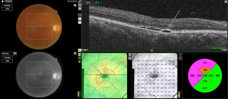
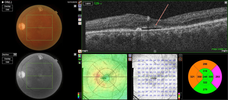
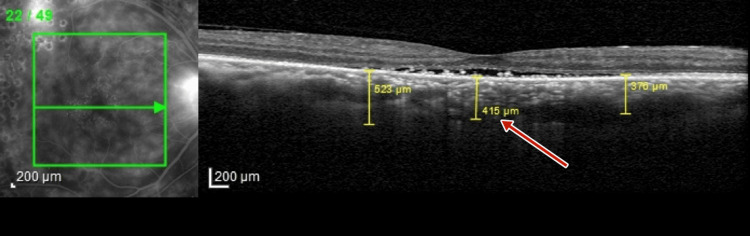
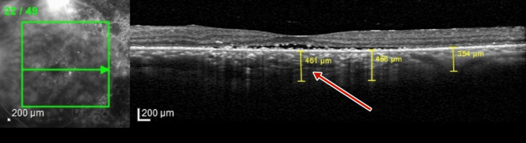
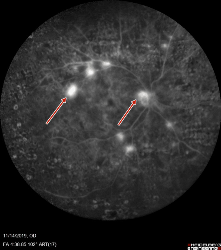
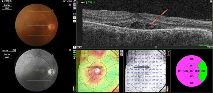
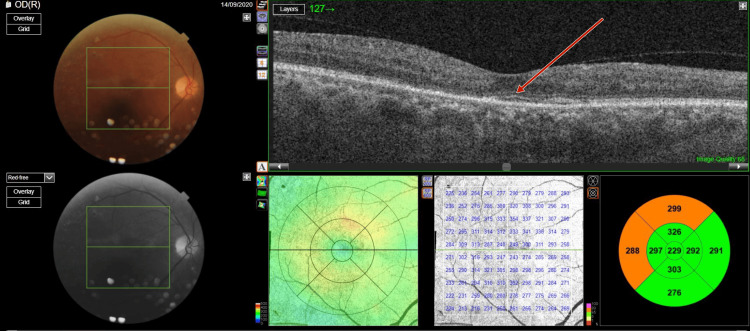
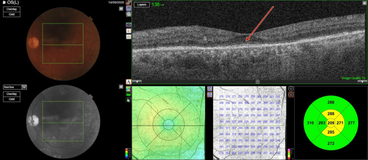
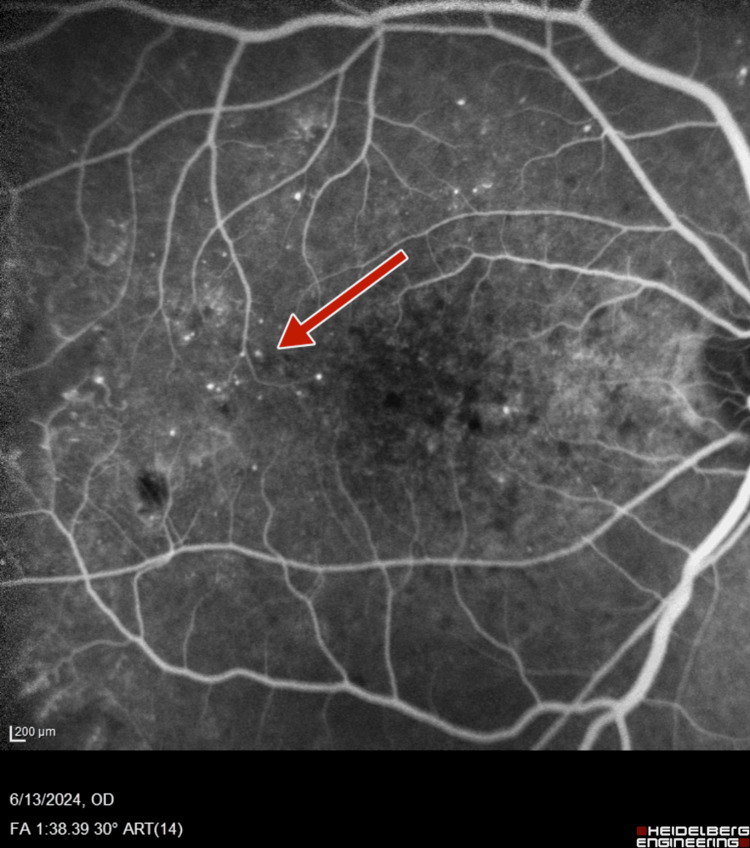
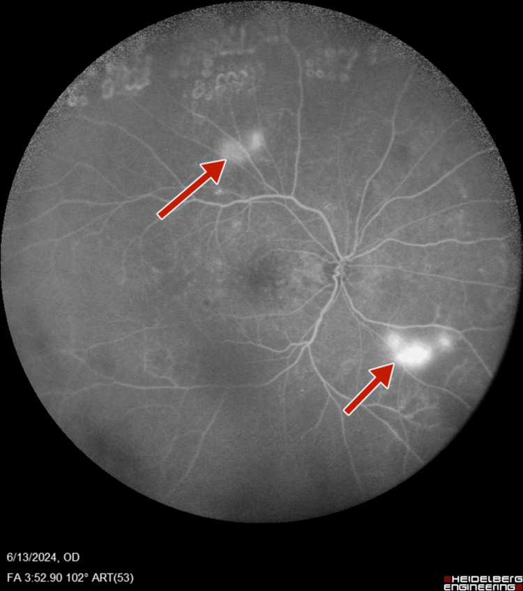
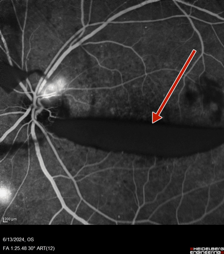
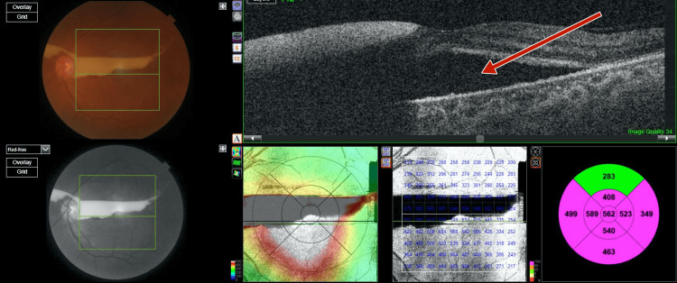
In this case series of two patients, we discuss pachychoroid as a risk factor for predicting exudative retinal detachment (RD) after panretinal photocoagulation (PRP). The first patient was a 55-year-old diabetic male with unstable proliferative diabetic retinopathy (PDR), serous pigment epithelial detachment (PED), and pachychoroid confirmed via fluorescein angiography (FA) and optical coherence tomography (OCT), who underwent PRP. Post-PRP, the patient complained of visual loss in both eyes. Subsequent FA and OCT confirmed the presence of exudative RD, which resolved after a course of non-steroidal anti-inflammatory eyedrops. The second patient was a 50-year-old male with PDR, serous PED, and pachychoroid confirmed via OCT, who underwent PRP. Post-PRP, he had reduced vision due to exudative RD. His vision improved upon the resolution of the exudative RD after three weeks. Pachychoroid is known to be associated with PDR. In the presence of pachychoroid, PRP-induced inflammation overwhelms the retinal pigment epithelium due to preexisting choroidal thickening, leading to exudative RD. These cases highlight how the identification of pachychoroid before laser PRP can help in predicting exudative RD as a post-procedure complication.

Keywords: exudative retinal detachment; laser complication; pachychoroid; panretinal photocoagulation laser; proliferative diabetic retinopathy (pdr).


Conflict of interest statement

Human subjects: Consent for treatment and open access publication was obtained or waived by all participants in this study. Conflicts of interest: In compliance with the ICMJE uniform disclosure form, all authors declare the following: Payment/services info: All authors have declared that no financial support was received from any organization for the submitted work. Financial relationships: All authors have declared that they have no financial relationships at present or within the previous three years with any organizations that might have an interest in the submitted work. Other relationships: All authors have declared that there are no other relationships or activities that could appear to have influenced the submitted work.

Figures

Figure 1
Figure 1. Photograph of right eye fundus and optical coherence tomography macula scan showing trace subretinal fluid with serous pigment epithelial detachment and normal foveal contour
Figure 2
Figure 2. Photograph of left eye fundus and optical coherence tomography macula scan showing trace subretinal fluid with serous pigment epithelial detachment and normal foveal contour
Figure 3
Figure 3. Pre-panretinal photocoagulation optical coherence tomography of right eye showing a subfoveal choroidal thickness of 415 µm suggestive of pachychoroid
Figure 4
Figure 4. Pre-panretinal photocoagulation optical coherence tomography of left eye showing a subfoveal choroidal thickness of 451 µm, suggestive of pachychoroid
Figure 5
Figure 5. Right-eye fluorescein angiography showing neovascularization of disc and retinal neovascularization along superotemporal arcade
Figure 6
Figure 6. Right-eye wide field fluorescein angiography showing neovascularization of disc and retinal neovascularization along superotemporal arcade
Figure 7
Figure 7. Left-eye fluorescein angiography showing neovascularization of disc and increased foveal avascular zone
Figure 8
Figure 8. Left-eye fluorescein angiography showing neovascularization of disc, retinal neovascularization, and increased foveal avascular zone
Figure 9
Figure 9. Right-eye post-panretinal photocoagulation optical coherence tomography showing exudative subretinal fluid at the fovea with hyperreflectivity suggestive of high protein content of the subretinal fluid
Figure 10
Figure 10. Left-eye post-panretinal photocoagulation optical coherence tomography showing exudative subretinal fluid at the fovea with hyperreflectivity suggestive of high protein content of the subretinal fluid
Figure 11
Figure 11. Right-eye post-panretinal photocoagulation fluorescein angiography suggestive of neovascularization of disc and retina
Figure 12
Figure 12. Right-eye post-panretinal photocoagulation wide-field fluorescein angiography suggestive of neovascularization of disc, as well as retina with staining at laser scars with increased foveal avascular zone
Figure 13
Figure 13. Left-eye post-panretinal photocoagulation fluorescein angiography suggestive of neovascularization of disc, with staining of the retinal blood vessels suggestive of loss of blood-retinal barrier
Figure 14
Figure 14. Left-eye post-panretinal photocoagulation wide-field fluorescein angiography suggestive of neovascularization of disc, with staining of the retinal blood vessels suggestive of loss of blood-retinal barrier
Figure 15
Figure 15. Follow-up right-eye optical coherence tomography scan showing complete resolution of subretinal fluid at macula
Figure 16
Figure 16. Follow-up left-eye optical coherence tomography scan showing complete resolution of subretinal fluid at macula
Figure 17
Figure 17. Right-eye optical coherence tomography showing trace subretinal fluid with serous pigment epithelial detachment with pachyhoroid
Figure 18
Figure 18. Left-eye optical coherence tomography showing trace subretinal fluid along with preretinal hemorrhage and incomplete posterior vitreous detachment
Figure 19
Figure 19. Right-eye fluorescein angiography showing microaneurysms
Figure 20
Figure 20. Right-eye late-phase fluorescein angiography showing retinal neovascularization
Figure 21
Figure 21. Left-eye fluorescein angiogram showing boat-shaped premacular hemorrhage
Figure 22
Figure 22. Left-eye late-phase fluorescein angiogram showing multiple retinal neovascularization
Figure 23
Figure 23. Right-eye optical coherence tomography showing subretinal fluid at macula due to exudative retinal detachment
Figure 24
Figure 24. Left-eye optical coherence tomography showing subretinal fluid at macula due to exudative retinal detachment

References

    1. Pachychoroid: an inherited condition? Lehmann M, Bousquet E, Beydoun T, Behar-Cohen F. Retina. 2015;35:10–16. - PubMed
    1. Pachychoroid spectrum disorders: an updated review. Brown R, Mohan S, Chhablani J. J Ophthalmic Vis Res. 2023;18:212–229. - PMC - PubMed
    1. Pattern scan laser photocoagulation: safety and complications, experience after 1301 consecutive cases. Velez-Montoya R, Guerrero-Naranjo JL, Gonzalez-Mijares CC, Fromow-Guerra J, Marcellino GR, Quiroz-Mercado H, Morales-Cantón V. Br J Ophthalmol. 2010;94:720–724. - PubMed
    1. Bullous exudative retinal detachment after retinal pattern scan laser photocoagulation in diabetic retinopathy. Nishikawa S, Kunikata H, Aizawa N, Nakazawa T. Case Rep Ophthalmol. 2017;8:475–481. - PMC - PubMed
    1. The role of diabetic choroidopathy in the pathogenesis and progression of diabetic retinopathy. Scuderi L, Fragiotta S, Di Pippo M, Abdolrahimzadeh S. Int J Mol Sci. 2023;24:10167. - PMC - PubMed

Publication types

LinkOut - more resources