[Preprint]. 2024 Dec 20:2024.11.27.625322.
doi: 10.1101/2024.11.27.625322.

Engineered CRISPR-Base Editors as a Permanent Treatment for Familial Dysautonomia

Affiliations

Engineered CRISPR-Base Editors as a Permanent Treatment for Familial Dysautonomia

Shuqi Yun et al. bioRxiv. .

Abstract

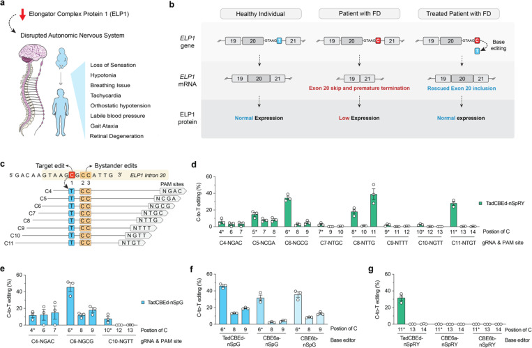
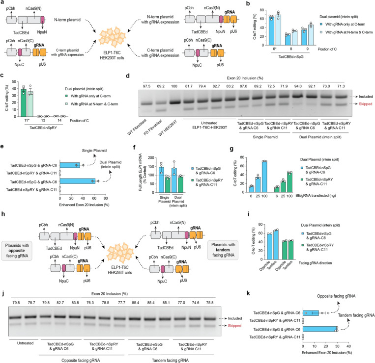
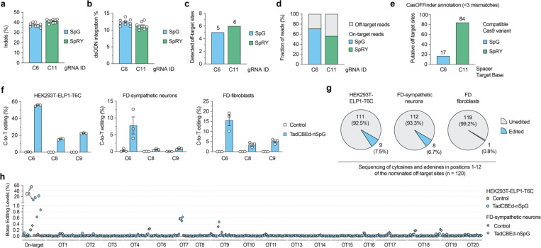
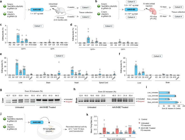
Familial dysautonomia (FD) is a fatal autosomal recessive congenital neuropathy caused by a T-to-C mutation in intron 20 of the Elongator acetyltransferase complex subunit 1 (ELP1) gene, which causes tissue-specific skipping of exon 20 and reduction of ELP1 protein. Here, we developed a base editor (BE) approach to precisely correct this mutation. By optimizing Cas9 variants and screening multiple gRNAs, we identified a combination that was able to promote up to 70% on-target editing in HEK293T cells harboring the ELP1 T-to-C mutation. These editing levels were sufficient to restore exon 20 inclusion in the ELP1 transcript. Moreover, we optimized an engineered dual intein-split system to deliver these constructs in vivo. Mediated by adeno-associated virus (AAV) delivery, this BE strategy effectively corrected the liver and brain ELP1 splicing defects in a humanized FD mouse model carrying the ELP1 T-to-C mutation and rescued the FD phenotype in iPSC-derived sympathetic neurons. Importantly, we observed minimal off-target editing demonstrating high levels of specificity with these optimized base editors. These findings establish a novel and highly precise BE-based therapeutic approach to correct the FD mutation and associated splicing defects and provide the foundation for the development of a transformative, permanent treatment for this devastating disease.

Keywords: CRISPR; Cas9; ELP1; bystander editing; genome editing; neurodegenerative disease; splicing.


Conflict of interest statement

Competing interests B.P.K., E.M. and C.R.R.A are inventors on a patent application filed by Mass General Brigham (MGB) that describes genome editing technologies to treat FD. B.P.K. and C.R.R.A. are inventors on additional patents or patent applications filed by MGB that describe genome engineering technologies, including to treat SMA. S.A.S. is an inventor on several U.S. and foreign patents and patent applications assigned to the Massachusetts General Hospital, including U.S Patents 8,729,025 and 9,265,766, both entitled “Methods for altering mRNA splicing and treating familial dysautonomia by administering benzyladenine,” filed on August 31, 2012 and May 19, 2014 and related to use of kinetin; and U.S. Patent 10,675,475 entitled, “Compounds for improving mRNA splicing” filed on July 14, 2017 and related to use of BPN-15477. E.M. and S.A.S. are inventors on an International Patent Application Number PCT/US2021/012103, assigned to Massachusetts General Hospital and entitled “ RNA Splicing Modulation” related to use of BPN-15477 in modulating splicing.B.P.K. is a consultant for EcoR1 capital, Novartis Venture Fund, and Jumble Therapeutics, and is on the scientific advisory boards of Acrigen Biosciences, Life Edit Therapeutics, and Prime Medicine. B.P.K. has a financial interest in Prime Medicine, Inc., a company developing therapeutic CRISPR-Cas technologies for gene editing. E.M. is a consultant for ReviR Therapeutics. C.R.R.A is a consultant for Ilios Therapeutics and Biogen and holds stocks in publicly traded companies developing gene therapies. B.P.K. and C.R.R.A interests were reviewed and are managed by MGH and MGB in accordance with their conflict-of-interest policies. The other authors declare no competing interests.

Figures

Figure 1.
Figure 1.. Development of cytosine base editors to correct the ELP1 T6C mutation causing FD.
a, Schematic of familial dysautonomia (FD) symptoms caused by reduced ELP1 protein levels affecting the autonomic nervous system. b, Schematic of FD caused by reduction in ELP1 protein levels due to a T-to-C (T6C) mutation in the position 6 of intron 20. c, Schematic of the genomic region surrounding the ELP1 T6C mutation, showing base editor guide RNA (gRNA) target sites; potential bystander edits are highlighted in orange boxes. d-e, C-to-T base editing to correct ELP1 T6C in homozygous HEK293T cells using TadCBEd, which comprises deaminase domains fused to PAM-variant SpCas9 enzymes nSpRY (d) and nSpG (e). Edited alleles were assessed via targeted sequencing and analyzed using CRISPResso2. f-g, Comparison of deaminase activity for correcting the ELP1 T6C mutation in homozygous HEK293T cells using TadCBEd, CBE6a, or CBE6b deaminase domains fused to SpG paired with gRNA C6 (f) or SpRY paired with gRNA C11 (g). Data in panels d-g from experiments in HEK 293T cells harboring the ELP1 T6C mutation; mean, s.e.m., and individual datapoints are shown for n = 3 independent biological replicates.
Figure 2.
Figure 2.. Engineering an intein-split system for in vivo delivery via adeno- associated virus (AAV).
a, Schematic of plasmid transfection experiments in HEK293T-ELP1-T6C cells using ITR-containing intein-split AAV production plasmid for TadCBEd fused with SpCas9 variants SpG or SpRY with N-terminus and C-terminus. Comparison between strategies with or without guide RNA (gRNA) expression in the N-term. b-c, C-to-T base editing to correct ELP1 T6C in homozygous HEK293T cells using AAV plasmids illustrated in panel a. d-e, ELP1 protein splicing assessing exon 20 inclusion and quantification for splicing enhancement in HEK293T ELP1 T6C cells treated with conventional base editor or intein-split plasmids. f, qPCR analysis of full-length ELP1 transcript expression following treatment with conventional and AAV plasmid systems using SpCas9 variants SpG paired with gRNA-C6 and SpRY paired with gRNA-C11). g, C-to-T editing efficiency using DNA titration of intein-split AAV plasmid in HEK293T ELP1 T6C cells. h, Schematic comparing opposite vs. tandem gRNA cassette orientation relative to SpCas9 expression in ITR-containing intein-split AAV production plasmids. i, C-to-T editing efficiency comparison between opposite and tandem gRNA orientations in intein-split AAV plasmids. j-k, ELP1 protein splicing assessing exon 20 inclusion and quantification for splicing enhancement when HEK293T ELP1 T6C cells treated with construct in panel h. Data in panels b-c, e-g, I, and k are from experiments in HEK293T cells; mean, s.e.m., and individual datapoints are shown for n = 2 or 3 independent biological replicates.
Figure 3.
Figure 3.. Base editing specificity in correcting the ELP1 T6C mutation.
a-b, Results from GUIDE-seq2 experiments including analysis of insertion or deletion (indels; panel a) and quantification of reads containing the GUIDE-seq2 dsODN tag (panel b) in ELP1 T6C HEK 293T cells when using SpCas9 variants SpG nuclease with gRNA-C6 or SpRY nuclease with gRNA-C11. c, Total number of GUIDE-seq2-detected off-target sites using SpG or SpRY. d, Percentage of total GUIDE-seq reads attributable to the on-target site or cumulative off-target sites. e, Number of putative off-target sites in the human genome with up to 3 mismatches for the spacers of gRNAs C6 and C11, as annotated by CasOFFinder. f, On-target C-to-T editing levels in HEK293T-ELP1-T6C cells, iPSC-derived FD-sympathetic neurons, and FD patient primary fibroblast cells when treated with TadCBEd-nSpG paired with gRNA-C6. g, Pie chart showing a summary of off-target sites validated for gRNA C6 and SpG across all 3 cell types in panel f, based on in silico CasOFFinder and GUIDE-seq2 nominations. h, Summary of on- and off-target base editing levels in three cell lines from panel f. Cells were untreated (naïve) or treated with TadCBEd-nPSG and gRNA C6. Genomic DNA was subjected to PCR for the on-target site and 19 off-target sites (nominated by CasOFFinder and GUIDE-seq2) with data analysis via CRISPResso2 for n = 3 independent biological replicates. For panels a-b and f, mean, standard errors of the mean (s.e.m.), and individual data points are shown for n = 3 or 9 independent biological replicates.
Figure 4.
Figure 4.. AAV-mediated delivery of base editors for in vivo ELP1 C6T editing.
a, Schematic of local retina injections of dual AAV2-BE vectors expressing intein-split TadCBEd-SpG and gRNA C6 into TgFD9 mice carrying the human ELP1 transgene with T6C mutation. Mice retina ganglion cells were isolated and sorted based on GFP expression at 4 or 6 weeks post-injection. b, Schematic of P1–2 intravenous (IV) injections of AAV9-BE vectors expressing intein-split TadCBEd-SpG and gRNA C6 into TgFD9 neonates. Mice were sacrificed, and tissues were harvested at 12 or 22 days post-injection. c-f, On-target C-to-T editing and potential bystander editing in the retina cells (c-d) or across different tissues (e-f) in each cohort treated base editors. g-h. ELP1 splicing in liver (g) and brain (h) tissues after AAV9-BE treatment. i, Quantification of exon 20 inclusion as percentage of the control in mice tissues are shown in panels g and h. j, Schematic of AAV2-BE transduction in iPSC-derived FD sympathetic neurons. Cell culture plates equipped with multielectrode arrays (MEAs) were used to record the electrical activity of sympathetic neurons. k, Electrical activity of iPSC-derived FD sympathetic neurons recorded at 5, 7, and 10 days post-transduction. Measurements were taken from both untreated and AAV2-BE-treated neurons. The mean firing rate was used to quantify overall neuronal firing activity. For panel k, mean, standard errors of the mean (s.e.m.), and individual data points are shown for n = 3-6 independent biological replicates.

References

    1. Slaugenhaupt S. A. et al. Tissue-specific expression of a splicing mutation in the IKBKAP gene causes familial dysautonomia. Am. J. Hum. Genet. 68, 598–605 (2001). - PMC - PubMed
    1. Slaugenhaupt S. A. Genetics of familial dysautonomia. Clin. Auton. Res. 12, I15–I19 (2002). - PubMed
    1. Cuajungco M. P. et al. Tissue-specific reduction in splicing efficiency of IKBKAP due to the major mutation associated with familial dysautonomia. Am. J. Hum. Genet. 72, 749–758 (2003). - PMC - PubMed
    1. Axelrod F. B. & Gold-von Simson G. Hereditary sensory and autonomic neuropathies: types II, III, and IV. Orphanet J. Rare Dis. 2, 39 (2007). - PMC - PubMed
    1. González-Duarte A., Cotrina-Vidal M., Kaufmann H. & Norcliffe-Kaufmann L. Familial dysautonomia. Clin. Auton. Res. Off. J. Clin. Auton. Res. Soc. 33, 269–280 (2023). - PubMed

Publication types

LinkOut - more resources