. 2025 Jan 31;99(1):e0165024.
doi: 10.1128/jvi.01650-24. Epub 2024 Dec 9.

The gE/gI complex is necessary for kinesin-1 recruitment during alphaherpesvirus egress from neurons

Affiliations

The gE/gI complex is necessary for kinesin-1 recruitment during alphaherpesvirus egress from neurons

Drishya Diwaker et al. J Virol. .

Abstract

Following reactivation of a latent alphaherpesvirus infection, viral particles are assembled in neuronal cell bodies, trafficked anterogradely within axons to nerve termini, and spread to adjacent epithelial cells. The virally encoded membrane proteins US9p and the glycoprotein heterodimer gE/gI of pseudorabies virus (PRV) and herpes simplex virus type 1 (HSV-1) play critical roles in anterograde spread, likely as a tripartite gE/gI-US9p complex. Two kinesin motors, kinesin-1 and kinesin-3, are implicated in the egress of these viruses, but how gE/gI-US9p coordinates their activities is poorly understood. Here, we report that PRV, in addition to associating with the kinesin-3 motor KIF1A, recruits the neuronal kinesin-1 isoforms KIF5A and KIF5C, but not the broadly expressed isoform KIF5B, during egress from differentiated CAD neurons. Similarly, in the axons of dorsal root ganglia (DRG)-derived sensory neurons, PRV colocalized with KIF5C but not KIF5B. In differentiated CAD cells, the association of KIF1A with egressing PRV was dependent upon US9p, whereas the recruitment of KIF5 isoforms required gE/gI. Consistent with these findings, the number of PRV particles trafficking within CAD neurites and the axons of DRG neurons increased when kinesin-1 motor activity was upregulated by hyperacetylating microtubules using trichostatin A (TSA) or tubacin, and this enhanced trafficking depended upon the presence of gE/gI. We propose that, following its recruitment by US9p, KIF1A delivers PRV particles to a location where KIF5 motors are subsequently added by a gE/gI-dependent mechanism. KIF5A/C isoforms then serve to traffic viral particles along axons, resulting in characteristic recrudescent infection.

Importance: Alphaherpesviruses include important human and veterinary pathogens that share a unique propensity to establish life-long latent infections in the peripheral nervous system. Upon reactivation, these viruses navigate back to body surfaces and transmit to new hosts. In this study, we demonstrate that the virus gE/gI-US9p membrane complex routes virus particles down this complex neuronal egress pathway by coordinating their association with multiple kinesin microtubule motors.

Keywords: alphaherpesviruses; anterograde transport; axonal transport; herpes simplex virus; pseudorabies virus.


Conflict of interest statement

G.A.S. is a co-founder of Thyreos, Inc., and serves on the scientific advisory board of EG427. G.A.S. has stock ownership in both entities.

Figures
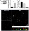
Fig 1
Fig 1
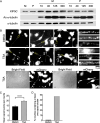
PRV recruits KIF5A and KIF1A to distinct populations of virus particles in differentiated CAD neurons. (A) Whole-cell lysates were prepared from equivalent numbers of P PRV-infected, undifferentiated CAD cells or differentiated CAD neurons (U-CAD and D-CAD, respectively) and then western blotted for KIF5A or β-actin as a loading control, as indicated at left. (B) Float-up fractions were prepared from P PRV-infected U-CAD and D-CAD cells, immunostained for KIF5A, and imaged for mCherry-tagged capsid fluorescence (red channel) and anti-KIF5A (blue channel). Red particles were scored for colocalization with KIF5A and plotted as a percent of total red particles in the field. Plotted values are mean and SD from the mean for 15,438 particles (U-CAD) and 17,321 particles (D-CAD), in each case counted from at least 15 microscopic fields. (C) Undifferentiated CAD cells were transfected to express mCitrine-KIF1A, differentiated, and infected with P PRV. Float-up fractions were prepared and immunostained for KIF5A then fields simultaneously imaged for mCherry-tagged capsids (red channel), mCitrine-KIF1A (green channel), and anti-KIF5A (blue channel). Red particles were scored for colocalization with mCitrine-KIF1A, KIF5A, or both motors simultaneously, as indicated on the X axis, and colocalization plotted as a percent of total red particles in the field. Plotted values are mean and SD from the mean for 27,995 particles counted from 25 microscopic fields. ****P ≤ 0.0001. (D) A microscopic field representative of those used to generate the data in (B). The red channel (mCherry-UL25p) and blue channel (anti-KIF5A) are shown in grayscale on the left, and a merged colored image are shown on the right (the anti-KIF5A image is pseudocolored in green). Merged panel scale bar: 20 µm. Five mCherry-UL25p fluorescing puncta that also exhibit anti-KIF5A fluorescence are indicated by arrows and labeled i–v. They are also shown at 7× higher magnification in the gallery below the merged field.
Fig 2
Fig 2
PRV recruits KIF5B and KIF1A to distinct populations of virus particles in undifferentiated CAD cells. (A) Whole-cell lysates were prepared from equivalent numbers of P PRV-infected U-CAD and D-CAD cells and then western blotted for KIF5B or β-actin as a loading control, as indicated at left. (B) Float-up fractions were prepared from P PRV-infected U-CAD and D-CAD cells, immunostained for KIF5B, and imaged for mCherry-tagged capsid fluorescence (red channel) and anti-KIF5B (blue channel). Red particles were scored for colocalization with KIF5B and plotted as a percent of total red particles in the field. Plotted values are mean and SD from the mean for 15,143 particles (U-CAD) and 28,002 particles (D-CAD) in each case counted from at least seven microscopic fields. (C) Undifferentiated CAD cells were transfected to express mCitrine-KIF1A and infected with P PRV. Float-up fractions were immunostained for KIF5B, and fields were simultaneously imaged for mCherry-tagged capsids (red channel), mCitrine-KIF1A (green channel), and anti-KIF5B (blue channel). Red particles were scored for colocalization with KIF1A, KIF5B, or both motors simultaneously, as indicated on the X axis, and colocalization plotted as a percent of total red particles in the field. Plotted values are mean and SD from the mean for 7,344 particles counted from five microscopic fields. (D) Schematic summarizing the data in Fig. 1 and 2. One or more enveloped PRV capsids reside within the bounding lipid envelope of a carrier vesicle, as indicated. In undifferentiated CAD cells (left), PRV recruits KIF1A to one population of carrier vesicles and the ubiquitous kinesin-1 KIF5B to another. Rarely are both motors observed on the same viral particle. A similar situation is seen in differentiated CAD neurons (right), except that PRV selects the neuron-specific kinesin-1 motors KIF5A and KIF5C, rather than KIF5B.
Fig 3
Fig 3
The consequences of US9p or gE/gI loss for recruitment of kinesins KIF1A, KIF5A, and KIF5C in differentiated CAD neurons. (A) Undifferentiated CAD cells were transfected to express mCitrine-KIF1A, differentiated, and infected with PRV strains P,ΔUS9p or ΔgE/gI, as indicated. Float-up fractions were prepared and red particles scored for colocalization with KIF1A (green channel) as in Fig. 1C. Colocalization is plotted as a percent of total red particles in the field. Plotted values are mean and SD from the mean for 61,923 P, 28,424 ΔUS9p, and 34,457 ΔgE/gI particles counted from 50, 43, and 48 microscopic fields, respectively. (B) Similar to (A) but float-up fractions were immunostained for KIF5A. Colocalization between red particles and KIF5A (blue channel) was plotted as a percent of total red particles in the field. Plotted values are mean and SD from the mean for 24,093 P, 24,472 ΔUS9p, and 23,418 ΔgE/gI particles, each counted from 18 microscopic fields. (C) Similar to (A) but float-up fractions were immunostained for KIF5C. Colocalization between red particles and KIF5C (blue channel) was plotted as a percent of total red particles in the field. Plotted values are mean and SD from the mean for 34,377 P, 30,216 ΔUS9p, and 41,692 ΔgE/gI particles, counted from 26, 25, and 29 microscopic fields, respectively. NS: not significant, **P ≤ 0.01 and ***P ≤ 0.001. (D) Schematic to summarize and explain these data. Egressing viral particles are represented as in Fig. 2D. The panel shows the predicted structure and properties of the P (upper left), ΔgE/gI (lower left), and ΔUS9p (upper right) strains of PRV. For the P virus, the bounding lipid bilayer of the carrier vesicle contains an intact gE/gI-US9p complex (represented by a red bar and blue lollipops, respectively). The deletion mutant viruses lack either US9p or gE/gI, as indicated, and the consequences of this for trafficking and kinesin recruitment are depicted. US9p and gE/gI are connected to the kinesins by broken lines to emphasize that the interaction between gE/gI-US9p and the motor proteins may not be direct. See text for more details.
Fig 4
Fig 4
The consequences of US9p or gE/gI loss for recruitment of kinesins KIF1A and KIF5B in undifferentiated CAD cells. (A) Undifferentiated CAD cells were transfected to express mCitrine-KIF1A then infected with PRV strain P,ΔUS9p or ΔgE/gI, as indicated. Float-up fractions were prepared and red particles scored for colocalization with KIF1A (green channel) as in Fig. 1C. Colocalization is plotted as a percent of total red particles in the field. Plotted values are mean and SD from the mean for 21,514 P, 20,735 ΔUS9p, and 23,805 ΔgE/gI particles, counted from 19, 18, and 22 microscopic fields, respectively. (B) Similar to (A) but float-up fractions were immunostained for KIF5B. Colocalization between red particles and KIF5B (blue channel) was plotted as a percent of total red particles in the field. Plotted values are mean and SD from the mean for 20,586 P, 27,202 ΔUS9p, and 29,171 ΔgE/gI particles, counted from 17, 23, and 29 microscopic fields, respectively.
Fig 5
Fig 5
PRV preferentially recruits KIF5C over KIF5B or KIF1A in the axons of DRG-derived primary sensory neurons. (A) Rat embryonic DRG-derived neurons were infected by PRV strain GS847, fixed between 0.5 and 18 h p.i. as indicated, then representative brightfield and red channel (mRFP1-capsid fluorescence) images merged. (B) Similar to (A) except neurons were infected by GS847 (left column) or its Δ(gE/gI-US9p)-derivative GS4384 (right column) then fixed at 24 h p.i. Infected neurons were imaged in brightfield (bottom row), for mRFP1-capsid fluorescence (middle row) or merged (top row). Scale bar: 10 µm. (C) DRG-derived neurons were infected by GS847, fixed at 18 h p.i., and immunostained for KIF5C. Each group of three images shows a representative length of infected axon imaged for anti-KIF5C fluorescence (green channel, bottom), GS847 mRFP1-capsid fluorescence (red channel, middle), or merged (top). Scale bar: 5 µm. Broken gray lines separates images collected from four individual axons. (D) DRG-derived neurons were infected exactly as in (C) and immunostained for KIF5B, or KIF5C, or imaged for mCitrine-KIF1A fluorescence expressed following infection by a recombinant lentivirus. Numbers of red-fluorescent capsid-associated axonal puncta colocalizing with each kinesin in a 32 mm length of infected axon were plotted as a percent of total puncta. Plotted values are the mean and SD from the mean for a total of 592 (mCitrine-KIF1A), 573 (anti-KIF5B), and 486 (anti-KIF5C) viral puncta, in all cases imaged in at least 20 axons in each of three independent experiments. Statistical significance was determined by one-way analysis of variance (ANOVA) with a post hoc Tukey test. (E) Identical experiment to (D) except that the intensity of mCitrine-KIF1A, anti-KIF5B, or anti-KIF5C fluorescence was measured in the soma and a 23 mm length of proximal axon. Plotted values are Log10 of the mean, and SD from the mean, for the sum of fluorescence intensity values recorded from the following numbers of soma and their proximal axons (soma/axons): mCitrine-KIF1A (8/16), KIF5B (10/20), and KIF5C (10/20).
Fig 6
Fig 6
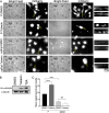
Effect of TSA on microtubule acetylation and PRV localization to CAD neurites. (A) Differentiated CAD neurons were infected with parental PRV (P) or mock infected (M) then incubated for 24 h in the absence of TSA (two left-hand lanes), or in the presence of 10, 40, 125, or 300 nM TSA, as indicated at top. Whole-cell lysates were prepared and western blotted for KIF5C, acetylated (AC)-α-tubulin or total α-tubulin, as indicated at left. (B) Top and middle row of images: P PRV-infected, differentiated CAD neurons were incubated with 125 nM TSA or dimethylsulfoxide (DMSO) solvent control (as indicated at left) for 24 h then fixed and imaged for mCherry-capsid fluorescence. Cell body fluorescence is deliberately overexposed in order to image particles in neurites. Regions boxed in broken yellow lines are magnified 5× at the right of each row, with those from the left, middle, and right panels depicted at top, middle, and bottom, respectively. White arrowheads indicate large viral particle clusters along the shafts of neurites. Bottom row of images: Identical experimental conditions to the middle row. Left pair of images and right pair of images represent two microscopic fields, each imaged for mCherry-capsid fluorescence or in bright field, as indicated. Arrowheads in the mCherry fields point to viral particle accumulations at the tips of neurites. Scale bars are 10 and 5 µm, as indicated. (C) Images similar to those in (B) were used to count numbers of individual capsid-associated viral particle puncta (other than those in clusters) present in infected neurites in DMSO- or TSA-treated CAD neurons. Plotted values are mean and SD from the mean for numbers of puncta counted in the neurites of 59 DMSO- and 57 TSA-treated, infected CAD neurons. (D) Images similar to those in (B) were used to determine the percentage of infected CAD neurons that contained at least one large capsid-associated cluster in their neurites, following DMSO or TSA treatment (99 and 96 infected CAD cell neurites examined, respectively).
Fig 7
Fig 7
Tubacin stimulates microtubule acetylation and gE/gI-dependent PRV accumulation in CAD neurites. (A) Differentiated CAD neurons were infected with P or ΔgE/gI PRV, then incubated for 24 h in the presence of 5 µM tubacin or DMSO solvent control (conditions are indicated at left of each row). Cells were then fixed and imaged in bright field or for mCherry-capsid fluorescence. Cell body fluorescence is deliberately overexposed in order to image particles in neurites. Two representative pairs of bright field and fluorescence images are shown in each row. Regions boxed in broken yellow lines are magnified 5× at the right of each row, with those from the left and right fluorescence panels depicted at the top and bottom, respectively. Scale bars are 10and 5 µm, as indicated. (B) Differentiated CAD neurons were infected with P PRV and incubated with 5 µM tubacin, 125 nM TSA or DMSO for 24 h then whole cell extracts prepared and western blotted for acetylated (AC)-α-tubulin or total α-tubulin, as indicated at left. (C) Images similar to those in (A) were used to count numbers of individual capsid-associated viral particle puncta (other than those in clusters) present in infected neurites in tubacin- or DMSO-treated CAD neurons. Plotted values are mean and SD from the mean for numbers of viral puncta counted in the neurites of 34 P-DMSO, 21 P-tubacin, 40 ΔgE/gI-DMSO, and 53 ΔgE/gI-tubacin treated CAD neurons.
Fig 8
Fig 8
Tubacin stimulates microtubule acetylation and gE/gI-dependent PRV accumulation in the axons of DRG-derived sensory neurons. DRG-derived neurons were infected with GS847 then treated with DMSO or 5 mM tubacin exactly as in Fig. 7. (A) Cells were fixed, permeabilized, and immunostained for acetylated α-tubulin. Ac-α-tubulin levels are elevated in tubacin treated neurons. Scale bar: 10 µm. (B) Numbers of fluorescent GS847 puncta present in the axons of tubacin, or DMSO-treated, DRG neurons. Plotted values are the mean and SD from the mean for 120 and 113 axons (tubacin- and DMSO-treated, respectively). (C) Similar to (B) but for PRV-GS4384 [Δ(gE/gI-US9p)]. Plotted values are the mean and SD from the mean for 61 and 73 axons (tubacin- and DMSO-treated, respectively).
Fig 9
Fig 9
Model for gE/gI-US9p-mediated kinesin recruitment during egress from sensory neurons. Schematic to explain and summarize our data. Egressing virus particles, the gE/gI-US9p complex, and kinesins are represented as in Fig. 3D. Microtubules are shown in green. PRV utilizes US9p to recruit KIF1A for traffic into the proximal axon, then at some later point exchanges KIF1A for KIF5A or KIF5C, using a gE/gI-dependent mechanism. KIF1A may be required for early stages of axonal traffic in sensory neurons because a high concentration of MAP2 in the proximal axon (gray rectangle) prevents kinesin-1 motors from associating with microtubules. See Discussion for more details.

References

    1. Smith G. 2012. Herpesvirus transport to the nervous system and back again. Annu Rev Microbiol 66:153–176. doi:10.1146/annurev-micro-092611-150051 - DOI - PMC - PubMed
    1. Diwaker D, Wilson DW. 2019. Microtubule-dependent trafficking of alphaherpesviruses in the nervous system: the ins and outs. Viruses 11:1165. doi:10.3390/v11121165 - DOI - PMC - PubMed
    1. Enquist LW, Husak PJ, Banfield BW, Smith GA. 1998. Infection and spread of alphaherpesviruses in the nervous system. Adv Virus Res 51:237–347. doi:10.1016/s0065-3527(08)60787-3 - DOI - PubMed
    1. Ambrosini AE, Borg KM, Deshmukh N, Berry MJ 2nd, Enquist LW, Hogue IB. 2024. Alpha herpesvirus exocytosis from neuron cell bodies uses constitutive secretory mechanisms, and egress and spread from axons is independent of neuronal firing activity. PLoS Pathog 20:e1012139. doi:10.1371/journal.ppat.1012139 - DOI - PMC - PubMed
    1. DuRaine G, Johnson DC. 2021. Anterograde transport of alpha-herpesviruses in neuronal axons. Virology (Auckl) 559:65–73. doi:10.1016/j.virol.2021.02.011 - DOI - PubMed

Publication types

MeSH terms

LinkOut - more resources