. 2024 Dec 9;20(12):e1012751.
doi: 10.1371/journal.ppat.1012751. eCollection 2024 Dec.

Mosquito-independent milk-associated transmission of zoonotic Wesselsbron virus in sheep

Affiliations

Mosquito-independent milk-associated transmission of zoonotic Wesselsbron virus in sheep

Marta Zimoch et al. PLoS Pathog. .

Abstract

Wesselsbron virus (WSLV) is a zoonotic, mosquito-borne orthoflavivirus endemic to sub-Saharan Africa, causing abortions and stillbirths in small ruminants. The life cycle of WSLV involves Aedes mosquitoes and various wildlife and domestic animals. Seminal studies in the 1950s have shown the zoonotic potential of WSLV, notably in accidental infections of laboratory workers exposed to infected material. More recent epidemiological studies suggest the emergence of clade I WSLV strains in peri-domestic and rural areas of western and eastern Africa. The pathobiology of recent clade I WSLV strains is unknown and no virus isolate is available. To address these gaps, we generated a recombinant clade I WSLV SA999 infectious clone (rSA999) by reverse genetics. Subsequently, lactating ewes were inoculated intravenously with the WSLV rSA999 strain or the clade II SAH177 strain in insect-free biocontainment stables. Inoculated ewes developed fever, viremia, and showed high levels of viral RNA at mucosal surfaces, and elevated viral titers in milk. Milk production was reduced, which directly affected the growth of the lambs, particularly within the rSA999 group. The ewes with higher WSLV titers in their milk in each group transmitted the infection to their lambs, which developed fever, prolonged viremia, and virus secretion. All infected animals produced high antibody titers with cross-neutralizing activity against both WSLV strains. Histopathology and blood biochemistry analysis indicated liver damage associated with necrotizing hepatitis lesions and active viral replication in some cases, which was more pronounced in the rSA999 group. Notably, only the SAH177-infected animals exhibited lesions consistent with meningoencephalitis, suggesting that WSLV clade II strains are neurotropic and that clade I strain are more hepatotropic. These findings demonstrate a previously unrecognized mode of vector-free transmission of WSLV that raises significant concerns for public and animal health.


Conflict of interest statement

The authors have declared that no competing interests exist.

Figures
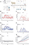
Fig 1
Fig 1. Wesselsbron disease kinetics in inoculated ewes and their lambs.
(A) Experimental design of the animal study. Lactating ewes and their lambs were randomly allocated to 3 groups and intravenously inoculated with 1ml of cell culture supernatant (mock), or 105 TCID50 of WSLV strains from clade I (rSA999) or clade II (SAH177). Daily monitoring and sampling included body temperature, clinical score, swabs, and blood samples from all animals, and milk collection from ewes and body weight of the lambs. (B,C) Body temperature (B) and clinical score (C) of ewes and lambs of the rSA999 group. (D,E) Body temperature (D) and clinical score (E) of ewes and lambs of the SAH177 group. (F) Body temperature of ewes and lambs of the mock group. (G) Relative body weight gain of the lambs of the 3 groups in percent of initial weight at day -1 prior to inoculation. Distinct symbols represent different animals throughout the manuscript within each group; lambs have the same symbol as their mother; twin lambs (SAH177 group) are distinguished by a dotted symbol. Differences between groups were analyzed by one-way ANOVA with Tukey post hoc test for comparing the area under the curve (AUC), and Mann-Whitney U test for daily individual differences in body weight. Underlined asterisks indicate statistical significance between groups showed in the same panel; asterisks next a group label indicates statistical significance compared to the mock group of the same age; blue asterisks indicate statistical differences between the rSA999 and the SAH177 groups; in (G) asterisks positioned on each day show statistical differences between rSA999 and mock (black asterisks) or with the SAH177 (blue asterisks) groups. p<0.05 was considered statistically significant (*p<0.05; **p<0.01; ***p<0.001; **** p<0.0001). Panel A was created with BioRender.
Fig 2
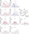
Fig 2. WSLV RNA load in blood, milk, and mucosal surfaces.
(A,B) Viremia determined by RT-qPCR in serum samples ewes and lambs of the rSA999 (A) and SAH177 (B) groups. (C,D) Viral RNA determined by RT-qPCR in milk of infected ewes of the rSA999 (C) and SAH177 (D) groups. (E) Infectious WSLV titers in milk samples at 5 dpi determined by end-point dilution and expressed as TCID50/ml. (F) Correlation analysis between RNA copies and infectious viral particles in the milk of infected ewes. (G-J) WSLV RNA copies in mucosal swabs from ewes: nasal (G), rectal (H), ocular (I), and oral (J). (K) WSLV RNA copies in oronasal swabs from lambs. Distinct symbols are used to represent different animals; within each group, lambs are denoted by the same symbol as their respective mother. Differences between groups were analyzed by one-way ANOVA with Tukey post hoc test for comparing the area under the curve (AUC). Underlined asterisks indicate statistical significance between groups showed in the same panel. p<0.05 was considered statistically significant (*p<0.05; **p<0.01; ***p<0.001; ****p<0.0001).
Fig 3
Fig 3. Neutralizing antibodies against WSLV during the course of infection.
(A,B) Neutralizing antibody titers in daily serum samples of the rSA999 (A) and SAH177 (B) groups. The mock group had no detectable neutralizing antibodies throughout the study. (C) Neutralizing antibody titers in milk at indicated time points. (D) Neutralizing serum antibody titers of indicated groups at 10 dpi against the heterologous virus. The neutralizing dose 50% (ND50) was determined using two-fold serial dilution of serum starting at 1:10 mixed with 200 FFU of WSLV. Distinct symbols are used to represent different animals; within each group, lambs are denoted by the same symbol as their respective mother. (A, B) Differences between groups were analyzed by one-way ANOVA with Tukey post hoc test for comparing the area under the curve (AUC). Underlined asterisks indicate statistical significance between groups showed in the same panel; blue asterisks indicate statistical differences in viremia between the rSA999 and the SAH177 groups. p<0.05 was considered statistically significant (****p<0.0001).
Fig 4
Fig 4. WSLV viral RNA loads in organs.
WSLV RNA levels were determined using RT-qPCR in the indicated organs of ewes and lambs collected at necropsy at 12 dpi, except for 1 ewe (red rhomboid) and its lamb (light red rhomboid), which were collected at 11 dpi as the lamb succumbed from the infection. The absolute WSLV RNA levels are expressed as copies per gram of tissue. Distinct symbols are used to represent different animals; within each group, lambs are denoted by the same symbol as their respective mother. No statistical analysis was performed.
Fig 5
Fig 5. Serum clinical biochemistry parameters in WSLV-infected sheep.
Serum samples collected daily were inactivated with 0.5% v/v NP-40 were analyzed for biomarker concentrations, including (A) aspartate aminotransferase (AST), (B) bilirubin, (C) bile acids, (D) adenosine deaminase, (E) cholesterol, (F) creatinine, and (G) blood urea nitrogen (BUN). The maximum and minimum values from the mock group of ewes and lambs were indicated for reference in gray shading background. (H) Group differences were analyzed by one-way ANOVA of the AUC with Tukey post hoc test. An asterisk on a specific group indicates differences compared to the age-matched mock; underlined asterisks indicate differences between infected groups. Statistical significance was considered for p<0.05 (***p<0.001; **** p<0.0001).
Fig 6
Fig 6. Histopathology and immunohistochemistry of WSLV NS1 in liver and brain samples.
(A,B) Semiquantitative scores of necrotizing hepatitis (liver) and non-suppurative meningoencephalitis (CNS) of ewes and lambs from rSA999 (A) and SAH177 (B) groups as none (0), 1 (mild), 2 (moderate) and 3 (severe). (C) Semiquantitative score of immunohistochemistry (IHC) positivity for WSLV NS1 in liver samples as negative (0), positive <5% of hepatocytes (1), 5–20% of hepatocytes (2) and >20% of hepatocytes (3). (D) Microscopic findings in the liver of animals infected with WSLV rSA999. (D1,3,5) H&E stains showing hepatic lesions of mild (D1), moderate (D3) and severe (D5) intensity. (D2,4,6) IHC for WSLV NS1 with low (D2), moderate (D4), and marked (D6) positivity. (D1) Ewe #2 (red square in panels 6A,C), multifocal hypereosinophilic, shrunken hepatocytes resembling Councilman bodies (arrows). (D2) Multifocal coarsely granular NS1 positivity is present within the cytoplasm of few hepatocytes (arrow). The hepatocytes show diffusely a mild intracytoplasmic vacuolar degeneration. (D3) Lamb #L2 (pink square in panels 6A,C), multifocal hypereosinophilic, shrunken hepatocytes with pyknosis and karyorrhexis (bold arrows), lymphocytic infiltrates (thin arrow) are scattered within the parenchyma. (D4) Multifocal coarsely granular NS1 positivity is present within the cytoplasm of hepatocytes (bold arrows) and Kupffer cells (thin arrow). (D5) Lamb #L5 (pink rhombus), there is diffuse loss of hepatic architecture, the hepatocytes show prominent intracytoplasmic vacuolar degeneration and multifocally are hypereosinophilic and fragmented (bold arrows). Inset: Round eosinophilic structure with marginated chromatin in the nucleus of a hepatocyte (intranuclear inclusion body) (bold arrow). (D6) Strong diffuse intracytoplasmic NS1 positivity in hepatocytes and Kupffer cells; the portal tract on the right of the image including its bile duct (asterisk) is negative. (E). Microscopic findings in H&E-stained CNS sections of animals infected with WSLV SAH177. (E1) Lamb #L8 (triangle in figure), cerebral cortex with accumulation of inflammatory leukocytes, predominantly lymphocytes, within the perivascular Virchow-Robin space (perivascular cuffs) (arrows) with multifocal neuronal degeneration and necrosis (thin arrows). (E2) Lamb #L8 (triangle in figure), brainstem with multifocal lymphocytic perivascular cuffs (bold arrows) adjacent to the lumen of the fourth ventricle (asterisk). (E3) Lamb #L10.1 (rhombus in figure), cerebral cortex with focal and prominent proliferation of glial cells (glial nodule, bold arrow) within the cortical grey matter. (E4) multifocal glial nodules within the white matter of the cervical spinal cord (bold arrows); the central canal of the spinal cord is on the top right of the image (asterisk).

Similar articles

Cited by

References

    1. Meganck RM, Baric RS. Developing therapeutic approaches for twenty-first-century emerging infectious viral diseases. Nat Med. 2021;27(3):401–10. Epub 20210315. doi: 10.1038/s41591-021-01282-0 . - DOI - PubMed
    1. Meurens F, Dunoyer C, Fourichon C, Gerdts V, Haddad N, Kortekaas J, et al.. Animal board invited review: Risks of zoonotic disease emergence at the interface of wildlife and livestock systems. Animal. 2021;15(6):100241. Epub 20210603. doi: 10.1016/j.animal.2021.100241 ; PubMed Central PMCID: PMC8172357. - DOI - PMC - PubMed
    1. Monge S, Garcia-Ortuzar V, Lopez Hernandez B, Lopaz Perez MA, Delacour-Estrella S, Sanchez-Seco MP, et al.. Characterization of the first autochthonous dengue outbreak in Spain (August-September 2018). Acta Trop. 2020;205:105402. Epub 20200220. doi: 10.1016/j.actatropica.2020.105402 . - DOI - PubMed
    1. Walsh MG, Sawleshwarkar S, Hossain S, Mor SM. Whence the next pandemic? The intersecting global geography of the animal-human interface, poor health systems and air transit centrality reveals conduits for high-impact spillover. One Health. 2020;11:100177. Epub 20201008. doi: 10.1016/j.onehlt.2020.100177 ; PubMed Central PMCID: PMC7543735. - DOI - PMC - PubMed
    1. Mackenzie JS, Gubler DJ, Petersen LR. Emerging flaviviruses: the spread and resurgence of Japanese encephalitis, West Nile and dengue viruses. Nat Med. 2004;10(12 Suppl):S98–109. doi: 10.1038/nm1144 . - DOI - PubMed