

Patients' Ability to Take Dermoscopic Follow-Up Images of Atypical Melanocytic Lesions With Smartphones: A Pilot Study
- PMID: 39652911
- PMCID: PMC11620195
- DOI: 10.5826/dpc.1404a268
Patients' Ability to Take Dermoscopic Follow-Up Images of Atypical Melanocytic Lesions With Smartphones: A Pilot Study
Abstract
Introduction: Short-term teledermoscopic monitoring helps to distinguish early melanomas from nevi. As the incidence of melanoma is increasing, there are several benefits of patients' taking their own dermoscopic images, but only a few previous studies have investigated the feasibility of this approach.
Objectives: To examine patients' ability to take evaluable dermoscopic images of atypical melanocytic lesions in need of short-term monitoring.
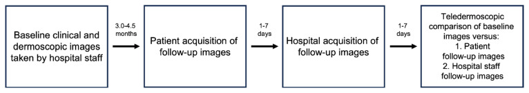
Methods: Patients were asked to take follow-up images in their homes using a borrowed dermoscope and their own smartphone. It was investigated whether the management decision differed when assessing follow-up images taken by patients compared to follow-up images taken by hospital staff. Lesions were rated as either changed, unchanged, or in need of further monitoring. In addition, image quality and patients' attitudes towards taking dermoscopic follow-up images were studied.
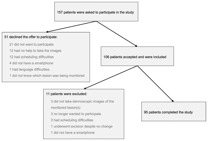
Results: Ninety-five patients with 132 lesions completed the study. Images taken by hospital staff were of better quality than images taken by patients (P<0.001). A total of 24 dermoscopic images taken by patients (18.2%) were of poor quality and considered unsuitable for assessment at follow-up. In the remaining 108 lesions, the management decision was concordant in 95 cases (88.0%). Most patients found the procedure to be easy to perform, and 76.0% of patients answered that they preferred self-photography.
Conclusions: Self-photography for teledermoscopic evaluation of atypical melanocytic lesions is feasible, but it results in worse image quality, which may lead to discordant evaluations. Dermoscopes used for this purpose need to be more user-friendly and maintain a higher technical standard.
Conflict of interest statement
Figures

References
-
- Menzies SW, Gutenev A, Avramidis M, Batrac A, McCarthy WH. Short-term digital surface microscopic monitoring of atypical or changing melanocytic lesions. Arch Dermatol. 2001;137(12):1583–9. DOI: - PubMed
LinkOut - more resources
Full Text Sources
Research Materials