. 2024 Dec 9;12(12):e009564.
doi: 10.1136/jitc-2024-009564.

DNA barcoded peptide-MHC multimers to measure and monitor minor histocompatibility antigen-specific T cells after allogeneic stem cell transplantation

Affiliations

DNA barcoded peptide-MHC multimers to measure and monitor minor histocompatibility antigen-specific T cells after allogeneic stem cell transplantation

Kyra J Fuchs et al. J Immunother Cancer. .

Abstract

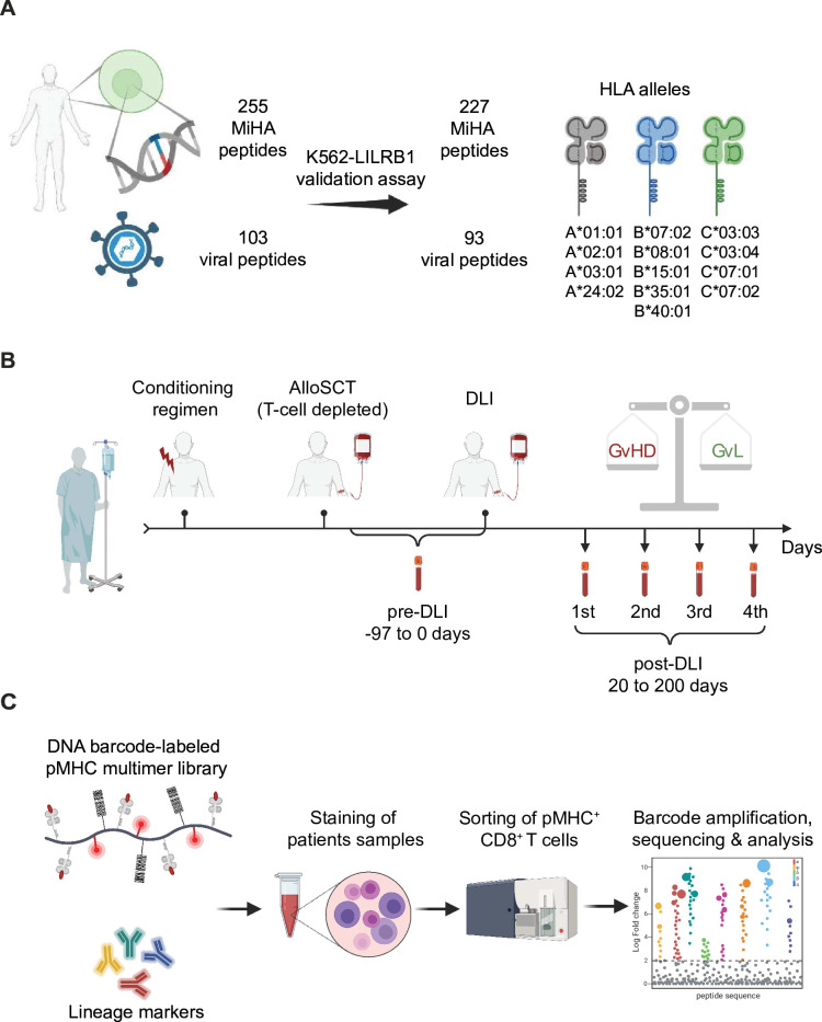
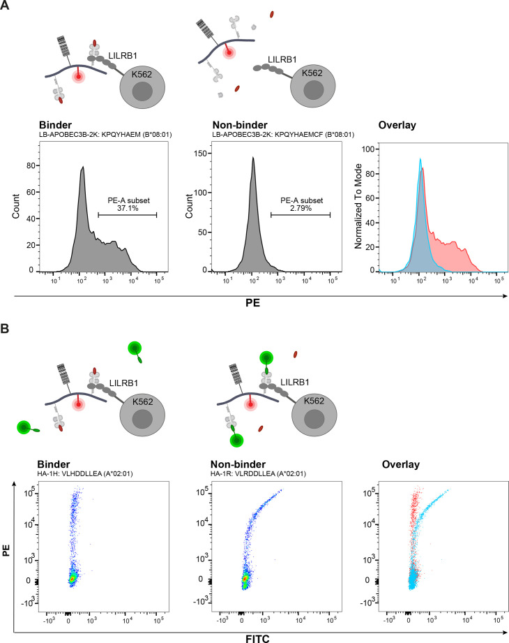
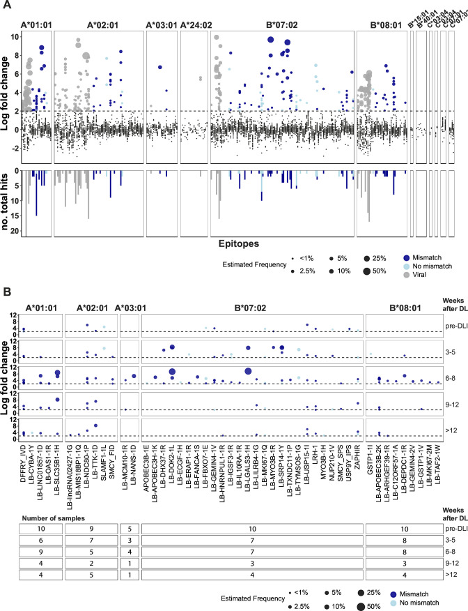
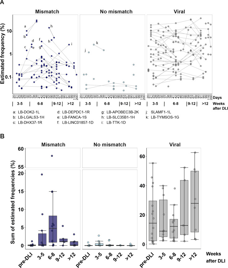
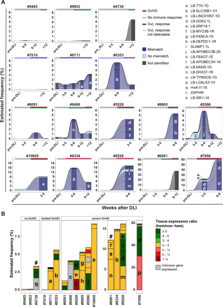
Allogeneic stem cell transplantation (alloSCT) provides a curative treatment option for hematological malignancies. After HLA-matched alloSCT, donor-derived T cells recognize minor histocompatibility antigens (MiHAs), which are polymorphic peptides presented by HLA on patient cells. MiHAs are absent on donor cells due to genetic differences between patient and donor. T cells targeting broadly expressed MiHAs induce graft-versus-leukemia (GvL) reactivity as well as graft-versus-host disease (GvHD), while T cells for MiHAs with restricted or preferential expression on hematopoietic or non-hematopoietic cells may skew responses toward GvL or GvHD, respectively. Besides tissue expression, overall strength of GvL and GvHD is also determined by T-cell frequencies against MiHAs.Here, we explored the use of DNA barcode-labeled peptide-MHC multimers to detect and monitor antigen-specific T cells for the recently expanded repertoire of HLA-I-restricted MiHAs. In 16 patients who experienced an immune response after donor lymphocyte infusion, variable T-cell frequencies up to 30.5% of CD8+ T cells were measured for 49 MiHAs. High T-cell frequencies above 1% were measured in 12 patients for 19 MiHAs, with the majority directed against mismatched MiHAs, typically 6-8 weeks after donor lymphocyte infusion and at the onset of GvHD. The 12 patients included 9 of 10 patients with severe GvHD, 2 of 3 patients with limited GvHD and 1 of 3 patients without GvHD.In conclusion, we demonstrated that barcoded peptide-MHC multimers reliably detect and allow monitoring for MiHA-specific T cells during treatment to investigate the kinetics of immune responses and their impact on development of GvL and GvHD after HLA-matched alloSCT.

Keywords: Graft versus host disease - GVHD; Graft versus leukemia; Hematologic Malignancies; T cell; Transplant.


Conflict of interest statement

Competing interests: None declared.

Figures

Figure 1
Figure 1. Overview of experimental setup. (A) A total of 255 MiHA peptides and 103 viral peptides were selected and evaluated for their ability to form stable pMHC-complexes with the K562-LILRB1 assay, resulting in 227 MiHA peptides and 93 viral peptides that were used for screening. (B) A cohort of 16 patients who underwent T cell-depleted alloSCT and DLI for treatment of hematological malignancies were screened. Samples before and after DLI, after which patients experienced an immune response defined by GvHD or conversion to full donor chimerism without GvHD, were analyzed. (C) Patient samples were stained with pMHC-multimer mixes and lineage markers to isolate pMHC-multimer-specific CD8+ T cells. Multimer mixes were specific for the HLA alleles as expressed by the patients. Barcodes from sorted pMHC-multimers were amplified, sequenced, and quantified to estimate peptide-specific T-cell frequencies. alloSCT, allogeneic stem cell transplantation; DLI, donor lymphocyte infusion; GvHD, graft-versus-host disease; GvL, graft-versus-leukemia; MiHA, minor histocompatibility antigen; pMHC, peptide-major histocompatibility complex.
Figure 2
Figure 2. Validation of peptides forming stable pMHC-complexes. (A) For pMHC-multimers that were produced by UV exchange, peptides were validated for their ability to form stable pMHC-complexes using K562 cells transduced with LILRB1, which binds to β2M as well as to the α3 domain of the MHC heavy chain. On addition, stable PE-labeled pMHC-complexes result in staining of LILRB1-transduced K562 cells. As examples, two peptide length variants are shown for LB-APOBEC3B-2K. Peptide KPQYHAEM binds to HLA-B*08:01 and leads to a stable pMHC-complex (left), whereas peptide KPQYHAEMCF does not bind and leads to an unstable pMHC-complex (middle). Both examples are also overlaid (right). (B) For pMHC-multimers that were produced using stabilized HLAs (HLA-A*02:01 and A*24:02), peptides were validated for stable binding in these pMHCs by adding a FITC-labeled peptide in the K562-LILRB1 assay. If the peptide stably binds in the pMHC, it cannot be outcompeted by the FITC-labeled peptide, leading to K562-LILRB1 cells that are exclusively stained with PE-labeled pMHC-complexes (left; HA-1 (VLHDDLLEA) stably binding to HLA-A*02:01). If the peptide does not stably bind, it is outcompeted by the FITC-labeled peptide leading to K562-LILRB1 cells that are stained with both PE-labeled and FITC-labeled pMHC-complexes (middle; allelic variant HA-1R (VLRDDLLEA) not stably binding). Overlay of both examples are displayed on the right. pMHC, peptide-major histocompatibility complex; PE, phycoerythrin.
Figure 3
Figure 3. Barcode-labeled pMHC-multimer detection of antigen-specific T cells in patients treated with alloSCT and DLI. (A) Overview of all DNA barcode measurements for samples from patients treated with alloSCT and DLI. Enriched barcodes were defined by a ≥2 log2 fold change and p<0.001 calculated using the Barracoda 1.8 pipeline. Enriched barcodes in the upper panel are colored for MiHAs that are mismatched (dark blue) or non-mismatched (light blue) in the respective patient-donor pairs, or for viral peptides (gray). Black data points with a log2 fold change <2 represent barcodes that were not significantly enriched (upper panel). Dot sizes represent estimated frequencies of the antigen-specific CD8+ T cells in the respective sample. The lower panel indicates the number of samples per barcode for which statistically significant enrichment was measured. (B) Enriched barcodes for MiHAs in alloSCT patients during treatment with DLI are shown for indicated time periods. All indicated antigens are validated MiHAs except for SLAMF1-1L, APOBEC3B-1E, MYO3B-1H, NUP210-1V, and GSTP1-1I, which are allelic variants of MiHAs. The table displays the number of samples included for indicated time periods. Within each time period, measurements for the sample with the highest total estimated T-cell frequency for MiHAs is shown for each patient. Multimers containing peptide length variants for MiHAs and allelic variants that may potentially bind to the same T cells were grouped, and the pMHC-multimer with the highest fold change within each group was selected per sample. alloSCT, allogeneic stem cell transplantation; DLI, donor lymphocyte infusion; MiHAs, minor histocompatibility antigens; pMHC, peptide-major histocompatibility complex.
Figure 4
Figure 4. Frequencies of MiHA-specific T cells in patient samples. (A) Indicated are estimated T-cell frequencies by pMHC-multimers with MiHAs that are mismatched (dark blue) or not mismatched (light blue) or pMHC-multimers with viral peptides (gray) in alloSCT patients after treatment with DLI. Data are shown for 11 patients with multiple samples after DLI screened by pMHC-multimers. In contrast to non-mismatched MiHAs, enriched barcodes for mismatched MiHAs and viral antigens were often detected in multiple samples after DLI indicated by connecting lines. (B) The sum of estimated T-cell frequencies is shown for all pMHC-multimers with mismatched or non-mismatched MiHAs or pMHC-multimers with viral peptides for each time period. Two patients were excluded as barcode sequence data were missing for post-DLI samples. alloSCT, allogeneic stem cell transplantation; DLI, donor lymphocyte infusion; MiHAs, minor histocompatibility antigens; pMHC, peptide-major histocompatibility complex.
Figure 5
Figure 5. Kinetics of MiHA-specific T cells in 16 patients who responded to DLI after HLA-matched alloSCT. (A) Estimated frequencies of MiHA-specific T cells by pMHC-multimer staining are shown for 16 patients who responded to DLI after HLA-matched alloSCT. Of the 16 patients, 3 patients responded to DLI without GvHD (patients #9465, #9953, #4739), 3 patients developed limited GvHD (patients #7010, #6711, #8353) and 10 patients had severe GvHD (#6091, #8490, #5528, #8905, #5596, #10605, #8334, #9528, #6061, #7956) after DLI. Each stack represents the estimated T-cell frequency for a MiHA that is mismatched (dark blue) or not mismatched (light blue) in the respective patient. For two samples (gray), frequencies of pMHC-multimer+ T cells measured during FACS sorting are displayed, but barcode sequence data were not determined due to PCR failure. Letters indicate MiHAs for which estimated T-cell frequencies of ≥1% of CD8+ T cells were detected. (B) Tissue expression of MiHAs targeted by T cells for the sample with the highest measured T-cell frequencies of each patient. Hashtags indicate two samples in which T-cell frequencies were measured by conventional pMHC-tetramer staining as barcode sequence data were missing. Colors indicate relative expression of the MiHA encoding gene in hematopoietic compared with non-hematopoietic cells using single cell RNA-Seq data of the Human Protein Atlas. alloSCT, allogeneic stem cell transplantation; DLI, donor lymphocyte infusion; GvHD, graft-versus-host disease; GvL, graft-versus-leukemia; MiHAs, minor histocompatibility antigens; pMHC, peptide-major histocompatibility complex.

Similar articles

References

    1. Biernacki MA, Sheth VS, Bleakley M. T cell optimization for graft-versus-leukemia responses. JCI Insight. 2020;5:e134939. doi: 10.1172/jci.insight.134939. - DOI - PMC - PubMed
    1. van der Zouwen B, Koster EAS, von dem Borne PA, et al. Feasibility, safety, and efficacy of early prophylactic donor lymphocyte infusion after T cell-depleted allogeneic stem cell transplantation in acute leukemia patients. Ann Hematol. 2023;102:1203–13. doi: 10.1007/s00277-023-05145-1. - DOI - PMC - PubMed
    1. Schmid C, Labopin M, Schaap N, et al. Long-term results and GvHD after prophylactic and preemptive donor lymphocyte infusion after allogeneic stem cell transplantation for acute leukemia. Bone Marrow Transplant. 2022;57:215–23. doi: 10.1038/s41409-021-01515-3. - DOI - PMC - PubMed
    1. Spierings E. Minor histocompatibility antigens: past, present, and future. Tissue Antigens. 2014;84:374–60. doi: 10.1111/tan.12445. - DOI - PubMed
    1. Griffioen M, van Bergen CAM, Falkenburg JHF. Autosomal Minor Histocompatibility Antigens: How Genetic Variants Create Diversity in Immune Targets. Front Immunol. 2016;7:100. doi: 10.3389/fimmu.2016.00100. - DOI - PMC - PubMed

MeSH terms

LinkOut - more resources