Computer Folding of Parallel DNA G-Quadruplex: Hitchhiker's Guide to the Conformational Space
- PMID: 39653644
- PMCID: PMC11628365
- DOI: 10.1002/jcc.27535
Computer Folding of Parallel DNA G-Quadruplex: Hitchhiker's Guide to the Conformational Space
Abstract
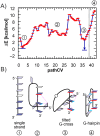
Guanine quadruplexes (GQs) play crucial roles in various biological processes, and understanding their folding pathways provides insight into their stability, dynamics, and functions. This knowledge aids in designing therapeutic strategies, as GQs are potential targets for anticancer drugs and other therapeutics. Although experimental and theoretical techniques have provided valuable insights into different stages of the GQ folding, the structural complexity of GQs poses significant challenges, and our understanding remains incomplete. This study introduces a novel computational protocol for folding an entire GQ from single-strand conformation to its native state. By combining two complementary enhanced sampling techniques, we were able to model folding pathways, encompassing a diverse range of intermediates. Although our investigation of the GQ free energy surface (FES) is focused solely on the folding of the all-anti parallel GQ topology, this protocol has the potential to be adapted for the folding of systems with more complex folding landscapes.
Keywords: DNA quadruplex; computational folding; enhanced sampling; kinetic partitioning mechanism; metadynamics; molecular dynamics; nudged elastic band; pathCV; transition path sampling.
© 2024 The Author(s). Journal of Computational Chemistry published by Wiley Periodicals LLC.
Conflict of interest statement
M.O. has a share in InSiliBio biosimulation company.
Figures


Similar articles
-
Folding of guanine quadruplex molecules-funnel-like mechanism or kinetic partitioning? An overview from MD simulation studies.Biochim Biophys Acta Gen Subj. 2017 May;1861(5 Pt B):1246-1263. doi: 10.1016/j.bbagen.2016.12.008. Epub 2016 Dec 13. Biochim Biophys Acta Gen Subj. 2017. PMID: 27979677 Review.
-
Computational Probing of the Folding Mechanism of Human Telomeric G-Quadruplex DNA.J Chem Inf Model. 2023 Oct 23;63(20):6366-6375. doi: 10.1021/acs.jcim.3c01257. Epub 2023 Oct 2. J Chem Inf Model. 2023. PMID: 37782649
-
Computational Insights into the Stability and Folding Pathways of Human Telomeric DNA G-Quadruplexes.J Phys Chem B. 2016 Jun 9;120(22):4912-26. doi: 10.1021/acs.jpcb.6b01919. Epub 2016 May 26. J Phys Chem B. 2016. PMID: 27214027
-
Mechanical Stability and Unfolding Pathways of Parallel Tetrameric G-Quadruplexes Probed by Pulling Simulations.J Chem Inf Model. 2024 May 13;64(9):3896-3911. doi: 10.1021/acs.jcim.4c00227. Epub 2024 Apr 17. J Chem Inf Model. 2024. PMID: 38630447 Free PMC article.
-
G-quadruplex dynamics.Biochim Biophys Acta Proteins Proteom. 2017 Nov;1865(11 Pt B):1544-1554. doi: 10.1016/j.bbapap.2017.06.012. Epub 2017 Jun 20. Biochim Biophys Acta Proteins Proteom. 2017. PMID: 28642152 Review.
References
-
- Sen D. and Gilbert W., “Formation of Parallel Four‐Stranded Complexes by Guanine‐Rich Motifs in DNA and Its Implications for Meiosis,” Nature 334 (1988): 364–366. - PubMed
-
- Ren J., Wang J., Han L., Wang E., and Wang J., “Kinetically Grafting G‐Quadruplexes Onto DNA Nanostructures for Structure and Function Encoding via a DNA Machine,” Chemical Communication 47 (2011): 10563–10565. - PubMed
-
- Phan A. T., “Human Telomeric G‐Quadruplex: Structures of DNA and RNA Sequences,” FEBS Journal 277 (2010): 1107–1117. - PubMed
-
- Karsisiotis A. I., O'Kane C., and Webba da Silva M., “DNA Quadruplex Folding Formalism—A Tutorial on Quadruplex Topologies,” Methods 64 (2013): 28–35. - PubMed
MeSH terms
Substances
Grants and funding
- 101092944/HORIZON EUROPE Framework Programme
- CA21101/European Cooperation in Science and Technology
- 90254/Ministry of Education, Youth and Sports of the Czech Republic
- CZ.02.01.01/00/22_008/0004587/Ministry of Education, Youth and Sports of the Czech Republic
- CZ.02.1.01/0.0/0.0/15_003/0000477/Ministry of Education, Youth and Sports of the Czech Republic
LinkOut - more resources
Full Text Sources
Miscellaneous

