

Phenotypic Spectrum of GNA11 R183C Mosaicism
- PMID: 39654261
- PMCID: PMC12118530
- DOI: 10.1111/pde.15802
Phenotypic Spectrum of GNA11 R183C Mosaicism
Abstract
Background: Many vascular anomalies harbor postzygotic somatic variants in GNAQ and GNA11; however, the phenotype of specific G-protein variants has not been well described. We report the clinical characteristics of 17 patients with a GNA11 R183C variant.
Methods: This case series is derived from a multinational cohort of vascular anomaly patients whose pathogenic mutations were identified using high-depth next generation sequencing. Data include vascular anomaly features, imaging reports, and extracutaneous manifestations of the GNA11 R183C variant.
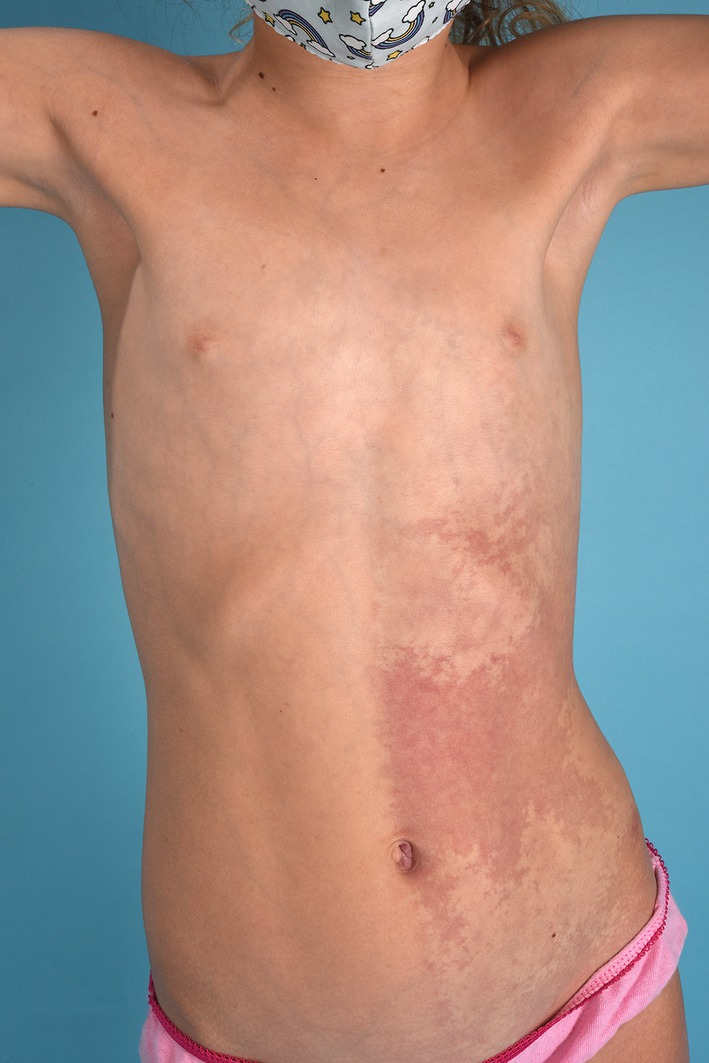
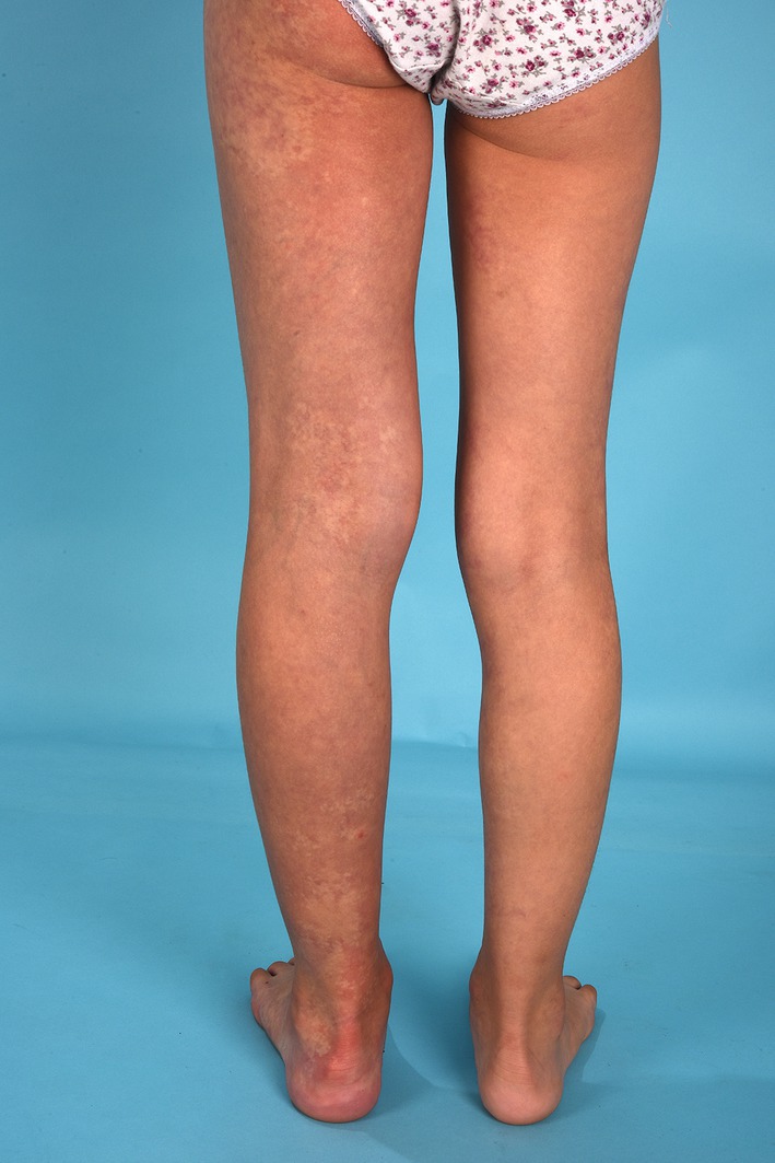
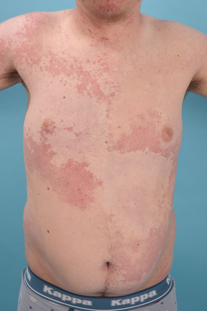
Results: We identified 17 subjects (median age 18 years [range 6-67]) with somatic GNA11 R183C variant. All patients had vascular lesions of the skin that presented as pink-to-red in children and deeper red in adults. Most lesions were large, poorly demarcated, and reticulated patches that were often bilaterally distributed. Nevus anemicus was observed in 53% (N = 9) and dermal melanocytosis in 13.3% (N = 2) of individuals. 82% (N = 14) of patients had limb growth discrepancies, and 1 patient had marked thoracic hypoplasia. 47% (N = 8) of patients had facial involvement, and 41% (N = 7) had forehead involvement. One patient experienced seizures due to right hemispheric leptomeningeal angiomatosis consistent with Sturge-Weber syndrome. Other findings included glaucoma (29%, N = 5) and psychomotor delay (29%, N = 5).
Conclusion: These findings contribute to our understanding of the clinical spectrum of GNA11 R183C capillary malformations (CMs); patients characteristically present with extensive, bilateral, poorly demarcated, pink-to-red CMs associated with nevus anemicus. Glaucoma and growth discrepancies (overgrowth or undergrowth) are common. Leptomeningeal angiomatosis and developmental delay can occur, appearing potentially less prevalent and severe than GNAQ-associated disease.
Keywords: genetic diseases; mechanisms; vascular malformation.
© 2024 The Author(s). Pediatric Dermatology published by Wiley Periodicals LLC.
Conflict of interest statement
Beth A Drolet is the Co‐founder and Board of Director at Arkayli Biopharma. Lisa M Arkin has no conflicts relevant to this study but is PI for research studies funded by Eli Lilly and Amgen, and receives consulting fees from Merck, Sanofi/Regeneron, and Nobel Pharma. DZ, LFSE, MI, EP, AJN, MMT, CEL, MM, NGO, SP, and EB have no conflicts to disclose.
Figures
References
-
- Tallman B., Tan O. T., Morelli J. G., et al., “Location of Port‐Wine Stains and the Likelihood of Ophthalmic and/or Central Nervous System Complications,” Pediatrics 87, no. 3 (1991): 323–327. - PubMed
-
- Enjolras O., Riche M. C., and Merland J. J., “Facial Port‐Wine Stains and Sturge–Weber Syndrome,” Pediatrics 76, no. 1 (1985): 48–51. - PubMed
Publication types
MeSH terms
Substances
Grants and funding
LinkOut - more resources
Full Text Sources
