

Polycythemia vera with acute coronary syndrome and bleeding as initial presentation: A case report and literature review
- PMID: 39654584
- PMCID: PMC11625114
- DOI: 10.1016/j.radcr.2024.10.109
Polycythemia vera with acute coronary syndrome and bleeding as initial presentation: A case report and literature review
Abstract
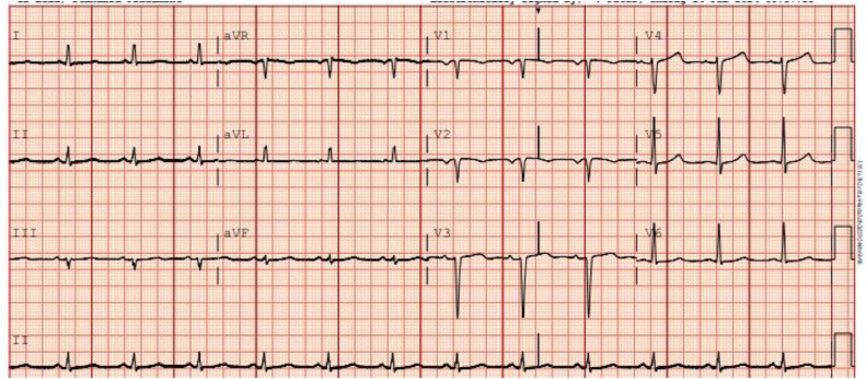
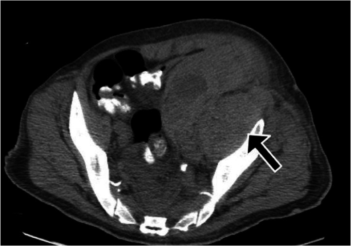
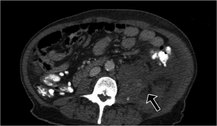
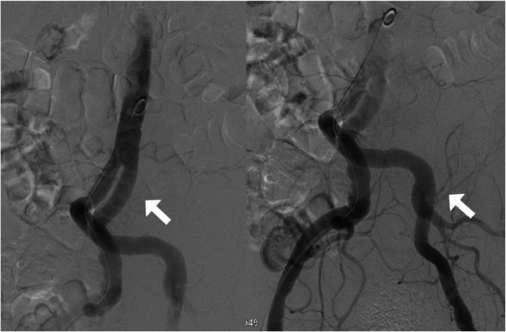
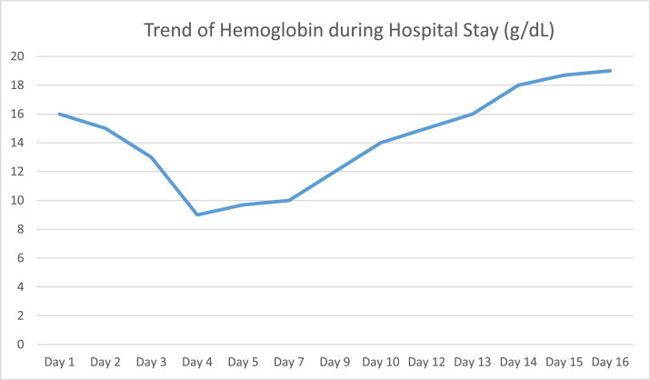
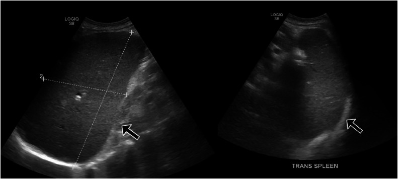
Polycythemia vera (PV) is a chronic myeloproliferative disorder characterized by increased red blood cell mass, leading to a heightened risk for thrombosis and hemorrhage. While thrombotic complications such as stroke, deep vein thrombosis, and pulmonary embolism are commonly associated with PV, coronary artery syndromes, as the initial presentation, are rare. Here, we present the case of a 73-year-old male who presented with severe chest pain and was diagnosed with non-ST-elevation myocardial infarction (NSTEMI). During his hospitalization, the patient experienced spontaneous psoas muscle hemorrhage, which prompted further investigation. Laboratory workup revealed elevated hemoglobin levels and a positive JAK2 V617F mutation, confirming a diagnosis of polycythemia vera. This case highlights the importance of considering myeloproliferative disorders in patients with atypical thrombotic and hemorrhagic events. It emphasizes the need for early diagnosis and appropriate treatment to optimize patient outcomes.
Keywords: Acute coronary syndrome; Myeloproliferative disorders; Polycythemia vera; Thromboembolic events.
© 2024 The Authors. Published by Elsevier Inc. on behalf of University of Washington.
Figures
References
-
- Stuart BJ, Viera AJ. Polycythemia vera. Am Fam Physician. 2004;69(9):2139–2144. - PubMed
-
- Adel G, Aoulia D, Amina Y, Aymen BA, Abdel-Hamid NM. Polycythemia vera and acute coronary syndromes: pathogenesis, risk factors and treatment. J Hematol Thromb Dis. 2013;1:107–112.
-
- Barbui T, Finazzi G, Falanga A. Myeloproliferative neoplasms and thrombosis. Blood. 2013;122:2176–2184. - PubMed
-
- Gruppo Italiano Studio Policitemia Polycythemia vera: the natural history of 1213 patients followed for 20 years. Gruppo Italiano Studio Policitemia. Ann Intern Med. 1995;123:656–664. - PubMed
Publication types
LinkOut - more resources
Full Text Sources
Miscellaneous
