. 2024 Nov 25:15:1484510.
doi: 10.3389/fmicb.2024.1484510. eCollection 2024.

Polymicrobial interactions influence Mycobacterium abscessus co-existence and biofilm forming capabilities

Affiliations

Polymicrobial interactions influence Mycobacterium abscessus co-existence and biofilm forming capabilities

Nishant Nandanwar et al. Front Microbiol. .

Abstract

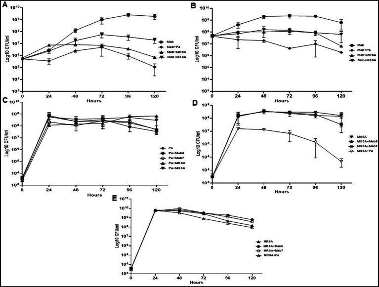
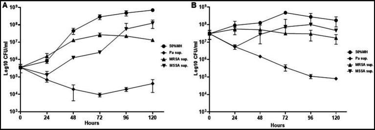
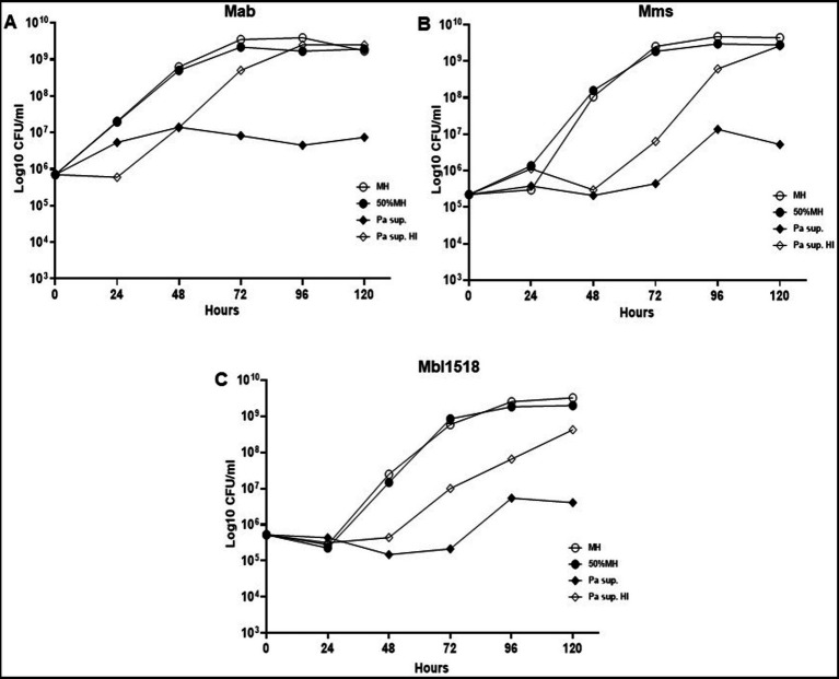
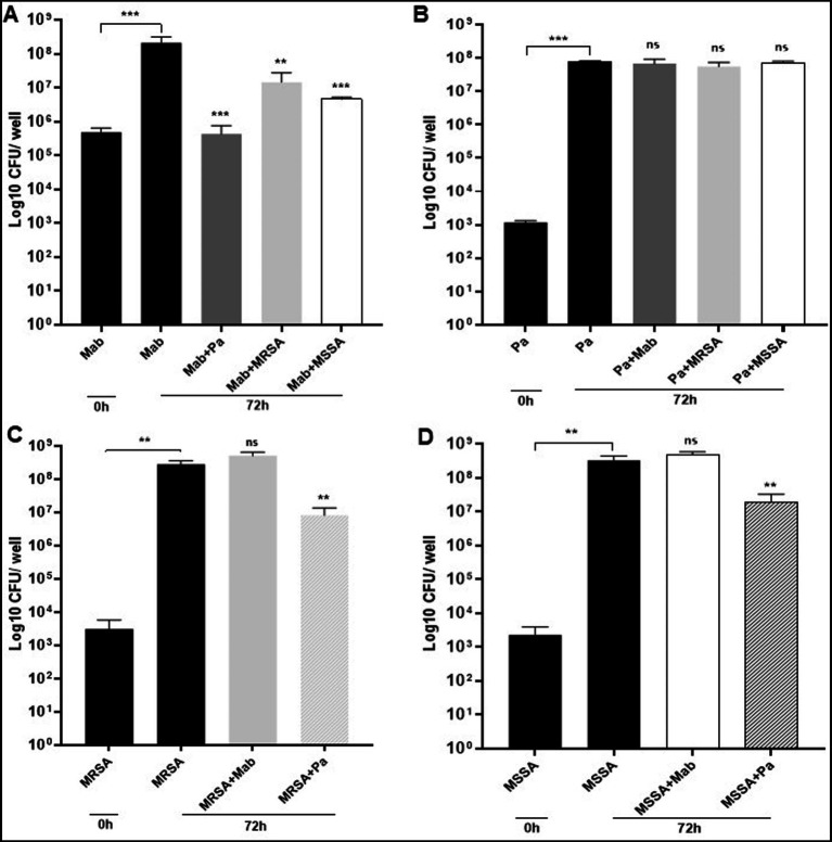
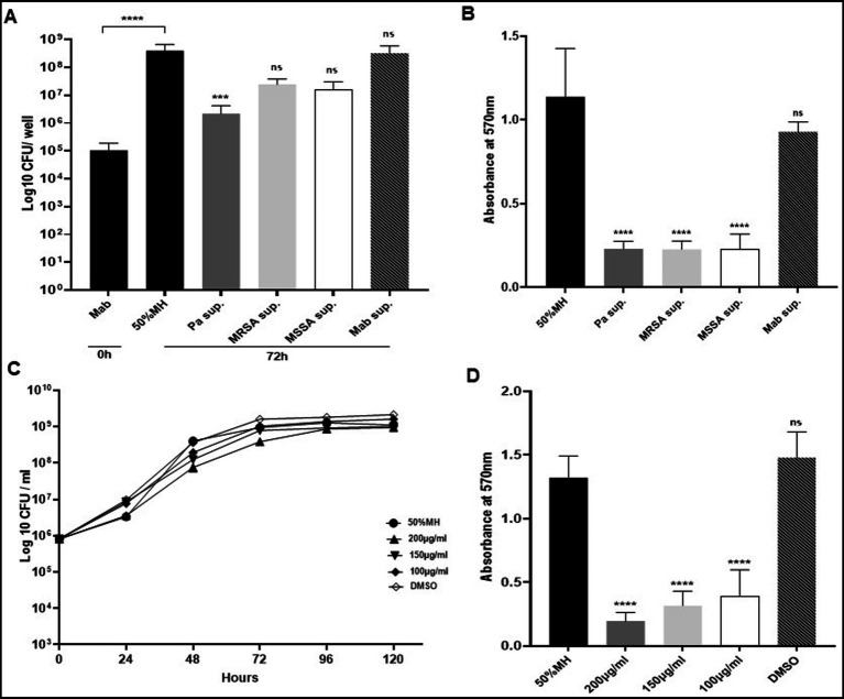
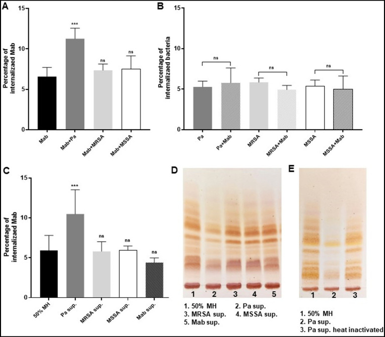
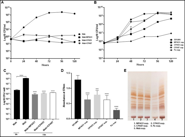
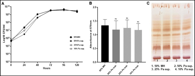
The lungs of patients with cystic fibrosis (CF) are vulnerable to persistent polymicrobial colonization by bacterial pathogens including Pseudomonas aeruginosa, Staphylococcus aureus, and the non-tuberculous mycobacterium (NTM) Mycobacterium abscessus. The polymicrobial milieu within the CF lung impacts individual species fitness, influences biofilm-forming capabilities, pathogenicity, production of virulence factors and even antimicrobial responses, all potentially compromising therapeutic success. Interaction studies among these CF pathogens are very limited, especially studies on the influences of P. aeruginosa and S. aureus on M. abscessus co-existence and virulence. Based on the little known thus far about coinfection of these pathogens, we hypothesize that the co-existence of P. aeruginosa and S. aureus alters M. abscessus virulence and phenotypic characteristics. We evaluated the direct (co-culture) and indirect (using supernatant) effects of P. aeruginosa and S. aureus on M. abscessus growth rate, biofilm formation, macrophage internalization and glycopeptidolipids (GPL) expression. Our observations indicate that P. aeruginosa and S. aureus exert a competitive behavior toward M. abscessus during direct contact or indirect interaction in-vitro, probably as is the case of polymicrobial infections in the lungs of patients with CF. This is the first report that demonstrates S. aureus inhibitory effects on M. abscessus growth and biofilm forming capabilities. Collectively, co-culture studies enhance our understanding of polymicrobial interactions during coinfection and can guide to establish better management of coinfections and treatment strategies for M. abscessus.

Keywords: Mycobacterium abscessus; Pseudomonas aeruginosa; Staphylococcus aureus; biofilm formation; co-culture; coinfection; polymicrobial interaction.


Conflict of interest statement

The authors declare that the research was conducted in the absence of any commercial or financial relationships that could be construed as a potential conflict of interest.

Figures

Figure 1
Figure 1
Growth pattern of Mab in co-culture (direct contact) with PaATCC and SA. (A) The presence of PaATCC and SA (collectively used for MRSA and MSSA) in co-culture with Mab (5 × 10⁵ CFU/ml) inhibited Mab growth when compared to the Mab monoculture. PaATCC in co-culture with Mab demonstrated the maximum inhibitory effect on Mab growth. (B) Even with the higher starting inoculum of Mab (5 × 10⁷ CFU/ml), the maximum growth inhibition of Mab was observed during co-culture with PaATCC. (C) On the contrary, the growth pattern of PaATCC was unaffected by the presence of Mab (higher and lower inoculum) and SA. (D,E) PaATCC also inhibited the growth of MSSA and MRSA in co-culture compared to the monoculture. However, both MRSA and MSSA were unaffected by the presence of Mab in co-culture. The starting inoculum for PaATCC, MRSA, and MSSA was 103 for each, respectively (Mab5, represents 5 × 10⁵ CFU/ml and Mab7, represents 7 × 10⁷ CFU/ml respectively). Growth curve experiments were performed in MH media as it supported the growth of all the participating bacteria in a co-culture. The graph represents the mean data and standard deviation of three independent experiments plated each time in duplicate. Pa represents PaATCC (please check Supplementary Tables S2A–C for statistical analysis of growth pattern of Mab, PaATCC, MSSA, and MRSA at different time points).
Figure 2
Figure 2
Growth pattern of Mab in supernatant (indirect contact) of PaATCC and SA. (A,B) Indirect contact of PaATCC, MRSA, and MSSA with Mab using supernatant prepared in MH media inhibited the growth of Mab (even at lower 5 × 10⁵ and higher 5 × 10⁷ CFU/ml of Mab starting inoculum). PaATCC supernatant demonstrated the maximum growth inhibitory effect on Mab growth. Supernatant prepared in MH media were diluted to 1:1 with MH media for the experiment and MH media diluted 1:1 with 1XPBS was used as a control for Mab growth (Pa sup. Represents PaATCC supernatant). The graph represents the mean data and standard deviation of three independent experiments plated each time in duplicate. Sup., supernatant (please check Supplementary Table S3 for statistical analysis of growth pattern of Mab under different conditions at different time points).
Figure 3
Figure 3
Heat inactivated supernatant does not inhibit M. abscessus growth. (A) The indirect contact of PaATCC inhibited Mab growth compared to the MH media control. However, the heat inactivated supernatant did not show growth inhibitory effect on Mab. (B,C) PaATCC supernatant also inhibited the growth of M. abscessus strains of other sub species such as the Mab. massiliense (Mms) and Mab. bolletii. 1518 (Mbl1518) compared to MH media control. However, the heat inactivated supernatant did not affect the growth of Mms and Mbl1518 (Pa sup. Represents PaATCC supernatant). The graph represents the mean data and standard deviation of two independent experiments plated each time in duplicate. Pa, PaATCC; sup., supernatant; HI, heat inactivated supernatant (please check Supplementary Table S4 for statistical analysis for A–C).
Figure 4
Figure 4
Mab biofilm forming capabilities in co-culture. (A) Mab biofilm forming capabilities were significantly altered or reduced in the presence of PaATCC, MRSA, and MSSA in co-culture after 72 h of incubation compared to the Mab monoculture. (B) The presence of Mab, MRSA, and MSSA in co-culture did not affect PaATCC biofilm forming capabilities after 72 h of co-culture. (C,D) Mab presence did not affect MRSA and MSSA biofilm. However, PaATCC in co-culture with MRSA and MSSA significantly reduced their biofilm forming capabilities after 72 h of incubation compared to the monoculture. The black solid bars represent controls that demonstrated significant biofilm formation by monoculture after 72 h of incubation compared to 0 h. The data represents mean with standard deviation from three independent experiments performed in triplicate each time. Statistics were performed using one-way ANOVA for multiple comparisons. Statistical significance is represented in figures by asterisks. ***p < 0.0005 and **p < 0.005. ns, non-significance; Pa, PaATCC.
Figure 5
Figure 5
Biofilm capabilities of Mab during indirect contact (using supernatant). (A) Mab biofilm forming capability was significantly reduced in the presence of PaATCC supernatant. Mab biofilm also reduced in presence of MRSA and MSSA supernatant but was not significant compared to the controls (Mab biofilm in diluted MH media and Mab supernatant respectively). Biofilm biomass by CFU counts was determined after 72 h of incubation. The black solid and black streaked bars represent controls that demonstrated significant biofilm formation after 72 h compared to 0 h (Pa sup. Represents PaATCC supernatant). (B) Biofilm forming capabilities as determined by CV staining of biofilm after 72 h revealed that Mab biofilm reduced significantly in presence of PaATCC, MRSA, and MSSA supernatant compared to the Mab biofilm in MH media and Mab supernatant used as controls. (C) Mab when grown in media supplemented with pyocyanin did not show any growth inhibition compared to the control (Mab grown in diluted MH media). (Equal volume of DMSO was used as a control as Pyocyanin was dissolved in DMSO) (please check Supplementary Table S5 for statistical analysis for C). (D) Pyocyanin significantly inhibited biofilm formation by Mab when compared to the media and DMSO controls. The growth curve data represents the mean and standard deviation of two independent experiments plated each time in duplicate. Biofilm forming assay was performed three times, each with at least six replicates each time. One-way ANOVA for multiple comparisons. Statistical significance is represented in figures by asterisks. ****p < 0.0001, ***p < 0.0005, ns, non-significance; Sup., supernatant.
Figure 6
Figure 6
Presence of PaATCC during coinfection enhances internalization of Mab in RAW264.7 macrophages. (A) Mab demonstrated enhanced and significant internalization in RAW264.7 macrophages when coinfected (direct contact) with PaATCC compared to the Mab alone infection. However, the coinfection of Mab with MRSA and MSSA did not affect the internalization of Mab in RAW macrophages. (B) Internalization of PaATCC, MRSA and MSSA during coinfection with Mab did not show significant difference compared to monoculture infection. (C) Mab grown in Pa supernatant (indirect contact) also demonstrated significant internalization in RAW264.7 macrophages compared to the controls (Mab grown in diluted MH media and Mab supernatant respectively). However, Mab cultured in MRSA and MSSA supernatant did not show any affect on Mab internalization in macrophages compared to the controls (Pa sup. Represents PaATCC supernatant). (D) Lipid isolation and TLC demonstrated lower expression of GPL in Mab grown in PaATCC supernatant compared to the controls (Mab grown in diluted MH media and Mab supernatant). MRSA and MSSA supernatant did not affect GPL expression in Mab. (E) Mab grown in heat inactivated PaATCC supernatant showed GPL expression compared to the normal PaATCC supernatant. All the infection data represents the mean and standard deviation of three independent experiments performed in triplicate each time. The TLC is the representative image of three different experiments. One-way ANOVA for multiple comparisons for infection assay and Student’s t test for comparing two groups was used. Statistical significance is represented in figures by asterisks. ***p < 0.0005. ns, non-significance.
Figure 7
Figure 7
Mab growth pattern, biofilm forming capabilities and GPL expression during direct and indirect contact with other PA. (A,B) Mab growth pattern of Mab during direct contact and in presence of supernatant from other PA isolates. Mab growth was significantly inhibited during direct contact but did not change much with supernatant of other PA isolates other than MPAO1 as compared to the Mab growth in PaATCC supernatant (shown here as a control) (Pa sup. Represents PaATCC supernatant) (please check Supplementary Tables S6, S7 for statistical analysis of A,B respectively). (C,D) Significant inhibition in the biofilm forming capacity of Mab was observed during direct and indirect contact of Mab with other PA isolates compared to Mab biofilm formed after 72 h in diluted MH media used as a control. (E) Growth of Mab in supernatants of other PA did not affect the GPL expression in Mab as compared to the control (Mab grown in Mab supernatant was taken as a control). The growth curve data represents the mean and standard deviation of two independent experiments plated each time in duplicate. Biofilm forming assay was performed three times, each with at least six replicates each time. TLC is the combined representative image of three different experiments. Statistics were performed using one-way ANOVA for multiple comparisons for biofilm assay. ****p < 0.0001, ***p < 0.0005. Sup., supernatant.
Figure 8
Figure 8
Mab revives its normal growth pattern, biofilm forming capabilities and GPL expression. Mab was grown in different concentrations (50, 25, and 10%) of PaATCC supernatant diluted with MH media and then centrifuged to pellet the bacteria. This pellet was resuspended in 1xPBS and used as an inoculum for growth curve, biofilm assay and for cultures to isolate GPL. Mab grown in diluted MH media (1:1 with 1xPBS) was used as a control. (A) Mab pretreated with PaATCC supernatant at different concentrations when grown in fresh media without any PaATCC supernatant revived its growth pattern comparable to the controlled growth condition (growth in diluted MH media) (please check Supplementary Table S8 for statistical analysis of A). (B) No significant difference was observed in the biofilm forming capabilities of Mab pretreated with PaATCC supernatant compared to the control (Mab grown in diluted MH media) after 120 h of incubation. (C) Lipid extraction and TLC of Mab grown in normal media after pretreatment with PaATCC supernatant did not show any significant difference in the expression of GPL as compared to the control (Mab grown in diluted MH media). The growth curve data represents the mean and standard deviation of three independent experiments plated each time in duplicate. The TLC is the representative image of three different experiments. Statistics were performed using one-way ANOVA for multiple comparisons. ns, non-significance; Sup., supernatant.

Similar articles

Cited by

References

    1. Archer N. K., Mazaitis M. J., Costerton J. W., Leid J. G., Powers M. E., Shirtliff M. E. (2011). Staphylococcus aureus biofilms: properties, regulation, and roles in human disease. Virulence 2, 445–459. doi: 10.4161/viru.2.5.17724, PMID: - DOI - PMC - PubMed
    1. Banks M. K., Bryers J. D. (1991). Bacterial species dominance within a binary culture biofilm. Appl. Environ. Microbiol. 57, 1974–1979. doi: 10.1128/aem.57.7.1974-1979.1991, PMID: - DOI - PMC - PubMed
    1. Beaudoin T., Yau Y. C. W., Stapleton P. J., Gong Y., Wang P. W., Guttman D. S., et al. . (2017). Staphylococcus aureus interaction with Pseudomonas aeruginosa biofilm enhances tobramycin resistance. NPJ Biofilms Microbiomes 3:25. doi: 10.1038/s41522-017-0035-0, PMID: - DOI - PMC - PubMed
    1. Bedrossian C. W., Donald Greenberg S., Singer D. B., Hansen J. J., Rosenberg H. S. (1976). The lung in cystic fibrosis. Hum. Pathol. 7, 195–204. doi: 10.1016/s0046-8177(76)80023-8 - DOI - PubMed
    1. Belardinelli J. M., Li W., Avanzi C., Angala S. K., Lian E., Wiersma C. J., et al. . (2021). Unique features of Mycobacterium abscessus biofilms formed in synthetic cystic fibrosis medium. Front. Microbiol. 12:743126. doi: 10.3389/fmicb.2021.743126, PMID: - DOI - PMC - PubMed

LinkOut - more resources