. 2024 Dec 3:19:12975-12998.
doi: 10.2147/IJN.S498099. eCollection 2024.

A Novel Triptolide Nano-Liposome with Mitochondrial Targeting for Treatment of Hepatocellular Carcinoma

Affiliations

A Novel Triptolide Nano-Liposome with Mitochondrial Targeting for Treatment of Hepatocellular Carcinoma

Lili Zhou et al. Int J Nanomedicine. .

Abstract

Background: Modern pharmacological studies have demonstrated that although triptolide (TP) is effective against hepatocellular carcinoma, it has poor water solubility and more toxic side effects. In this study, we used triptolide (TP), a bioactive constituent in Tripterygium wilfordii Hook F, as a model drug to develop a novel nano-liposome drug delivery system for the treatment of liver tumours.

Methods: We constructed a functionally-modified triptolide liposome (FA+TPP-TP-Lips) using the film-dispersion method and investigated its physicochemical properties, mitochondrial targeting of hepatic tumour cells, in vitro and in vivo anti-hepatic tumour activity and its mechanism.

Results: The prepared FA+TPP-TP-Lips had a particle size of 99.28 ± 5.7 nm, a PDI of 0.20 ± 0.02, a zeta potential of 1.2 ± 0.08 mV, and an encapsulation rate of 74.37% ± 1.07%.FA+TPP-TP-Lips facilitates the cellular uptake of drug delivery systems and improves their targeted delivery to mitochondria. The results of cell efficacy showed that FA+TPP-TP-Lips significantly inhibited the growth of liver cancer cells, decreased mitochondrial membrane potential, and increased intracellular ROS, thus enhancing the highest apoptosis rate of liver cancer cells. The targeted liposomes (FA-TP-Lips, TPP-TP-Lips, and FA+TPP-TP-Lips) had some degree of inhibitory migration effect on Huh-7 cells relative to the unmodified TP-Lips. Studies on tumor-bearing mice demonstrated that FA+ TPP-TP-Lips effectively accumulated in tumor tissues and significantly inhibited the growth of subcutaneous tumors, achieving a tumor inhibition rate of 79.37%. FA+ TPP-TP-Lips demonstrated an enhanced anti-liver tumor effect and significantly mitigated the hepatotoxicity and systemic toxicity associated with TP.

Conclusion: In summary, the results of this study can provide a feasible solution for improving the mitochondrial targeting of nano-liposomes, and lay a foundation for further developing a novel nano targeting preparation of triptolide for the treatment of hepatocellular carcinoma.

Keywords: Folic acid; Hepatocellular Carcinoma; Mitochondrial Targeting; Triphenylphosphine ion; Triptolide.


Conflict of interest statement

The authors declare that they have no known competing financial interests or personal relationships that could have appeared to influence the work reported in this paper.

Figures

Scheme 1
Scheme 1
Synthesis of FA+TPP-TP-Lips and its anti-hepatic tumour effects.
Figure 1
Figure 1
Characterisation of liposomes.(A)Transmission electron micrographs of liposomes.(B) Size distributions.(C) Zeta potential.
Figure 2
Figure 2
Haemolytic assessment of liposomes.(A) TP-Lips; (B) TPP-TP-Lips; (C) FA-TP-Lips; (D) FA+TPP-TP-Lips.
Figure 3
Figure 3
Inhibitory effect of liposomes on Huh-7 and AML12 cell proliferation (n = 3). (A) Cell inhibition rate of FA+TPP-TP-Lips at different action times. (B) IC50 values of different experimental groups acting on Huh-7 cell line. Data are expressed as the mean±SD (n=3), (C) IC50 values of different experimental groups acting on AML12 cell line. Data are expressed as the mean±SD (n=3). *p < 0.05, ** p < 0.01, **** p < 0.0001.
Figure 4
Figure 4
Effects of liposomes on Huh-7 cell migration (n = 3). (A) Comparative graph of Huh-7 cell migration; and (B) Migration rate of Huh-7 cells.
Figure 5
Figure 5
Uptake of liposomes modified with different ligands by Huh-7 cells.(A) Free C6; (B)C6- Lips; (C) FA-C6- Lips; (D) TPP-C6- Lips; (E) FA+TPP-C6- Lips.
Figure 6
Figure 6
Mitochondrial morphology of Huh-7 cells after treatment with liposomes modified with different ligands (n=3). (A) Free TP; (B) TP-Lips; (C) FA-TP-Lips; (D) TPP-TP- Lips; (E) FA+TPP-TP-Lips.
Figure 7
Figure 7
Effects of liposome intervention on mitochondrial membrane potential (n = 3). (A) Microscopic images of mitochondrial membrane potential; (B) Histogram of the fluorescence intensity ratio of JC-1 aggregate/JC-1 monomer.Data are expressed as the mean±SD (n=3). **p < 0.01, ****p < 0.0001.
Figure 8
Figure 8
Effect of different liposomal interventions on ROS levels in Hun-7 cells (n = 3). (A) Quantification of ROS levels in Hun-7 cells using flow cytometry. (B) Statistical analysis of ROS levels. Data are expressed as the mean±SD (n=3). ns, not significant, ***p < 0.001, ****p < 0.0001.
Figure 9
Figure 9
Effect of liposome treatment on apoptosis of Huh-7 cells (n = 3).(A) Control; (B) Free TP; (C) TP-Lips; (D) FA-TP-Lips; (E) TPP-TP-Lips; (F) FA+TPP-TP-Lips. (G) Quantitative data of apoptosis rate of Huh-7 cells. Data are expressed as the mean±SD (n=3). (H) Quantitative data of necrosis rate of Huh-7 cells. Data are expressed as the mean±SD (n=3). ns, not significant. ***p < 0.001, ****p < 0.0001.
Figure 10
Figure 10
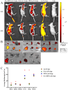
Fluorescence images of (A) in vivo and (B) in vitro hormonal mice after treatment with different therapeutic methods. (C) The quantitative data of average radiance (p/s/cm2/sr) in each organ.
Figure 11
Figure 11
In vivo antitumour effects. (A) Tumors collected on day 12 (B) Tumor volume growth curve (C) Tumor weight. Data are expressed as the mean±SD (n=5). (D) H&E staining of tumor sections (E) Tumor proliferation index of BALB/C nude mice bearing tumor. Data are expressed as the mean±SD (n=3). (F) Apoptosis rate of tumor in BALB/C nude mice. Data are expressed as the mean±SD (n=3). *p < 0.05, **p < 0.01, ***p < 0.001, ****p < 0.0001, ns, not significant.
Figure 12
Figure 12
Safety evaluation of liposomes. (A) H&E staining of the organs of Homer BALB/C nude mice (n=3). (B) Results of biochemical indexes of the liver and kidney in each group of Homer BALB/C nude mice (n= 3). (C) Change rate of body weight in BALB/C nude mice during the administration cycle (n=5).

Similar articles

References

    1. Zheng Y, Niu X, Xue W, et al. The Role of Alternative Splicing Factors hnRNP G and Fox-2 in the Progression and Prognosis of Esophageal Cancer. Dis Markers. 2022;2022:3043737. doi:10.1155/2022/3043737 - DOI - PMC - PubMed
    1. Ueshima E, Nishiofuku H, Takaki H, et al. Hepatic Artery Embolization Induces the Local Overexpression of Transforming Growth Factor β1 in a Rat Hepatoma Model. Liver Cancer. 2020;9(1):63–72. doi:10.1159/000502774 - DOI - PMC - PubMed
    1. Yu G, Ali Z, Sajjad Khan A, et al. Preparation, Pharmacokinetics, and Antitumor Potential of Miltefosine-Loaded Nanostructured Lipid Carriers. Int J Nanomed. 2021;16:3255–3273. doi:10.2147/IJN.S299443 - DOI - PMC - PubMed
    1. Chen ZW, Hu J-F, Wang Z-W, et al. Circular RNA circ-MTHFD1L induces HR repair to promote gemcitabine resistance via the miR-615-3p/RPN6 axis in pancreatic ductal adenocarcinoma. J Exp Clin Cancer Res. 2022;41(1):153. doi:10.1186/s13046-022-02343-z - DOI - PMC - PubMed
    1. Fialho MFP, Brum ES, Becker G, et al. Kinin B(2) and B(1) Receptors Activation Sensitize the TRPA1 Channel Contributing to Anastrozole-Induced Pain Symptoms. Pharmaceutics. 2023;15(4):1136. doi:10.3390/pharmaceutics15041136 - DOI - PMC - PubMed

MeSH terms