. 2024 Nov 30:2024:8027006.
doi: 10.1155/bmri/8027006. eCollection 2024.

Induction of Innate Immune Memory in LPS-Primed Microglial Cells by Water-Soluble Chitosan

Affiliations

Induction of Innate Immune Memory in LPS-Primed Microglial Cells by Water-Soluble Chitosan

Vo Thuy Anh Thu et al. Biomed Res Int. .

Abstract

Innate immune memory or trained immunity refers to a long-lasting response of the innate immune cells against repeated exposure to the homogenous or heterogenous infectious agent. The trained immunity is induced through epigenetic modification and is characterized by the change of both intracellular immunological signaling and cellular metabolism. Recently, different groups have tried to establish protocols to generate trained innate immune cells. However, the molecular basis of innate memory induction remains poorly understood. Here, we evaluated the impact of water-soluble chitosan on the innate immune memory induction in microglial cells primed with LPS. The trained-immune response was accessed by measuring proinflammatory markers, metabolic change, and epigenetic modification. We showed that the stimulation/restimulation with LPS only caused a robust reduction of iNOS, and proinflammatory cytokines, indicating induced immune tolerance. In contrast, the treatment of chitosan induces long-lasting memory microglial cells accompanied by a high level of iNOS, increased lactate production, induced epigenetic modification, and the upregulation of proinflammatory cytokines upon further exposure to the same stimulus. These findings suggest that chitosan induces microglial-trained immunity by targeting distinct epigenetic and metabolic pathways; therefore, chitosan treatment may provide a novel approach for targeting innate immunity towards a memory-like response in an in vitro model.

Keywords: microglia; trained immunity; water-soluble chitosan.


Conflict of interest statement

The authors declare no conflicts of interest.

Figures
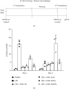
Figure 1
Figure 1
Enhancement of microglial reactivity by WSC upon secondary LPS exposure. (a) Experimental timeline: depicts the sequence of initial exposure, rest period, and restimulation to evaluate the effects of WSC on microglial activation following a second LPS challenge. (b) Gene expression analysis: quantitative real-time PCR results showing mRNA levels after the first (24 h postinitial exposure) and second stimulations (6 h post-re-exposure). Results were normalized against a DMSO control to assess changes in gene expression indicative of microglial activation or priming by WSC. ⁣p < 0.05 and ⁣∗∗p < 0.005 versus DMSO.
Figure 2
Figure 2
Long-term modulation of microglial reactivation by WSC post-LPS exposure. (a) Experimental design timeline: outlines the sequence of LPS and/or WSC treatment, followed by washout periods and subsequent restimulation to investigate the lasting impact of WSC on microglial activation. (b) Gene expression evaluation: quantitative real-time PCR of iNOS mRNA levels after initial treatment and 6 h post-restimulation. The comparative analysis against a DMSO baseline control highlights shifts in gene expression that indicate alterations in microglial activation and potential priming effects of WSC. ⁣p < 0.05 and ⁣∗∗p < 0.005 versus DMSO; #p < 0.05 and ##p < 0.005.
Figure 3
Figure 3
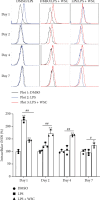
WSC amplifies microglial iNOS response to secondary LPS stimulation. Microglial cells were initially treated with LPS (100 ng/mL) alone or with 0.02% WSC for 24 h, followed by washout periods of 1, 3, or 6 days. iNOS expression was measured using flow cytometry after both initial treatment and 12 h post-restimulation, assessing WSC's impact on microglial activation. ⁣p < 0.05 and ⁣∗∗p < 0.01 versus DMSO; #p < 0.05 and ##p < 0.005.
Figure 4
Figure 4
WSC modulates LPS-induced epigenetic factor expression in human microglia. Microglial cells were treated with LPS (100 ng/mL) alone or with 0.02% WSC for 24 h, followed by washout periods of 1, 3, or 6. mRNA expression of histone methyltransferases (a) SETD1B and (b) SUV39H1 was measured by qPCR after initial stimulation and 6 h post-restimulation. ⁣p < 0.05 and ⁣∗∗p < 0.005 versus DMSO; #p < 0.05 and #p < 0.001.
Figure 5
Figure 5
Influence of WSC on LPS-induced lactate production in human microglia. Microglial cells were exposed to LPS (100 ng/mL) alone or with 0.02% WSC for 24 h, followed by 3 or 6 washouts. (a) LDHA mRNA expression was measured after the first 24-h stimulation 6 h restimulation. (b) The concentration of lactate in the culture supernatant was determined using a lactate fluorometric assay kit after both the first and second 24-h stimulation periods. ⁣p < 0.05 and ⁣∗∗p < 0.005 versus DMSO; #p < 0.05 and ##p < 0.005.
Figure 6
Figure 6
WSC augments microglial activation in response to LPS. Microglial cells were subjected to LPS (100 ng/mL) alone or with 0.02% WSC for a 6-h, followed by a 24-h washout and restimulation for 6 h. After restimulation, cells were fixed with formaldehyde, permeabilized with Triton X-100, and incubated with an anti-Iba1 antibody. (a) Fluorescence signals were captured using a fluorescent microscope at 200x magnification, with scale bars representing 100 μm. (b) The corrected total cell fluorescence (CTCF) was quantitatively assessed using ImageJ software, providing a measure of Iba1 expression levels indicative of microglial activation. ⁣p < 0.01; ns denotes nonsignificant differences.
Figure 7
Figure 7
WSC amplifies proinflammatory cytokine expression in microglia following LPS challenge. Microglial cells were treated with LPS (100 ng/mL) alone or with 0.02% WSC for 24 h, followed by a washout and restimulation with the same treatments. (a) mRNA level of IL-1β (a) and TNF-α (b) were measured via qPCR after the first 24-h stimulation and 6 h post-restimulation. ⁣p < 0.05 and ⁣∗∗p < 0.005 versus DMSO; #p < 0.05, ##p < 0.005, and ###p < 0.001.
Figure 8
Figure 8
Conceptual model of enhanced microglial immune response to LPS via trained immunity induced by WSC. The model depicts three distinct phenotypic responses: endotoxin tolerance (yellow line): represents a reduced inflammatory response to repeated LPS exposure, marked by decreased cytokine production, which is commonly referred to as endotoxin tolerance. naïve inducible response (red line): illustrates the typical acute inflammatory response of naïve microglial cells to initial LPS exposure, marked by significant cytokine secretion and activation. Trained immunity (blue line): demonstrates the enhanced microglial response to a second LPS challenge in the presence of WSC, characterized by increased cytokine production, epigenetic modifications, and metabolic reprogramming.

Similar articles

References

    1. Xing Z., Afkhami S., Bavananthasivam J., et al. Innate immune memory of tissue-resident macrophages and trained innate immunity: re-vamping vaccine concept and strategies. Journal of Leukocyte Biology . 2020;108(3):825–834. doi: 10.1002/JLB.4MR0220-446R. - DOI - PubMed
    1. Liu Y., Liang S., Ding R., et al. BCG-induced trained immunity in macrophage: reprograming of glucose metabolism. International Reviews of Immunology . 2020;39(3):83–96. doi: 10.1080/08830185.2020.1712379. - DOI - PubMed
    1. Hamon M. A., Quintin J. Innate immune memory in mammals. Seminars in Immunology . 2016;28(4):351–358. doi: 10.1016/j.smim.2016.05.003. - DOI - PubMed
    1. Netea M. G., Domínguez-Andrés J., Barreiro L. B., et al. Defining trained immunity and its role in health and disease. Nature Reviews Immunology . 2020;20(6):375–388. doi: 10.1038/s41577-020-0285-6. - DOI - PMC - PubMed
    1. Iwanowycz S., Wang J., Altomare D., Hui Y., Fan D. Emodin bidirectionally modulates macrophage polarization and epigenetically regulates macrophage memory. Biological Chemistry . 2016;291(22):11491–11503. doi: 10.1074/jbc.M115.702092. - DOI - PMC - PubMed

LinkOut - more resources