Higher density of CD4+ T cell infiltration predicts severe renal lesions and renal function decline in patients with diabetic nephropathy
- PMID: 39654881
- PMCID: PMC11625791
- DOI: 10.3389/fimmu.2024.1474377
Higher density of CD4+ T cell infiltration predicts severe renal lesions and renal function decline in patients with diabetic nephropathy
Abstract
Background: More evidence have shown that the combination of immune and inflammatory mechanism was critical in diabetic nephropathy (DN). However, the relationship between CD4+ T cells and the development of DN is still unclear. Therefore, this study will focus on this issue from the perspective of clinicopathology.
Methods: From September 2019 to December 2022, a total of 112 adult patients with DN were enrolled in the study. According to the density of CD4+ T cell infiltration based on immunostaining, the patients were divided into high-CD4 group (56 patients) and low-CD4 group (56 patients). Another 25 diabetic patients with minimal change disease (non-diabetic nephropathy, NDN) was reviewed as control group in clinical and molecular analysis. The clinical parameters, morphological features, and molecular characteristics were compared. The predictive value of CD4+ T cells for DN prognosis was also investigated.
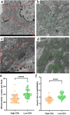
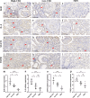
Results: DN patients in the high-CD4 group suffered from higher proteinuria and lower estimated glomerular filtration rate (eGFR) level than those in the low-CD4 group and NDN patients. Renal biopsy in the high-CD4 group presented with more severe glomerular lesions, higher density of interstitial inflammation, and more severe tubular atrophy/interstitial fibrosis than in the low-CD4 group. Multivariate logistic analysis indicated that the density of CD4+ T cell infiltration could independently predict the severity of tubular atrophy/interstitial fibrosis. In addition, more severe mitochondrial damage of renal tubular epithelial cells and a more obvious expression of Bcl6, IL-6, STAT3, and TGFβ1 were observed in DN patients of the high-CD4 group, indicating the possible mechanism of CD4+ T cells involving the progression of DN. Multivariate Cox regression analysis revealed that a higher intensity of interstitial CD4+ T cell deposition remained as an independent predictor of the double endpoint with doubling of baseline serum creatinine or end-stage renal disease.
Conclusion: The high density of CD4+ T cell infiltration was associated with renal function decline and severity of renal lesions and predicted poor renal survival for DN patients.
Keywords: CD4+ T cells; clinicopathological analysis; diabetic nephropathy; interstitial fibrosis; tubular atrophy.
Copyright © 2024 Han, Xu, Li, Lei, Li, Zhao and Liu.
Conflict of interest statement
The authors declare that the research was conducted in the absence of any commercial or financial relationships that could be construed as a potential conflict of interest.
Figures


Similar articles
-
The association of hematuria on kidney clinicopathologic features and renal outcome in patients with diabetic nephropathy: a biopsy-based study.J Endocrinol Invest. 2020 Sep;43(9):1213-1220. doi: 10.1007/s40618-020-01207-7. Epub 2020 Mar 9. J Endocrinol Invest. 2020. PMID: 32147762
-
Tissue expression of tubular injury markers is associated with renal function decline in diabetic nephropathy.J Diabetes Complications. 2017 Dec;31(12):1704-1709. doi: 10.1016/j.jdiacomp.2017.08.009. Epub 2017 Aug 24. J Diabetes Complications. 2017. PMID: 29037450
-
Renal histology in diabetic nephropathy predicts progression to end-stage kidney disease but not the rate of renal function decline.BMC Nephrol. 2020 Jul 18;21(1):285. doi: 10.1186/s12882-020-01943-1. BMC Nephrol. 2020. PMID: 32682403 Free PMC article.
-
Lessons learned from studies of the natural history of diabetic nephropathy in young type 1 diabetic patients.Pediatr Endocrinol Rev. 2008 Aug;5 Suppl 4:958-63. Pediatr Endocrinol Rev. 2008. PMID: 18806710 Review.
-
Macrophage Phenotype and Fibrosis in Diabetic Nephropathy.Int J Mol Sci. 2020 Apr 17;21(8):2806. doi: 10.3390/ijms21082806. Int J Mol Sci. 2020. PMID: 32316547 Free PMC article. Review.
Cited by
-
Integrating bioinformatics and machine learning to identify glomerular injury genes and predict drug targets in diabetic nephropathy.Sci Rep. 2025 May 15;15(1):16868. doi: 10.1038/s41598-025-01628-5. Sci Rep. 2025. PMID: 40374840 Free PMC article.
References
MeSH terms
LinkOut - more resources
Full Text Sources
Medical
Research Materials
Miscellaneous

