. 2024 Nov 25:14:1459810.
doi: 10.3389/fcimb.2024.1459810. eCollection 2024.

Galectin-3 plays a key role in controlling infection by Toxoplasma gondii in human trophoblast cells and human villous explants

Affiliations

Galectin-3 plays a key role in controlling infection by Toxoplasma gondii in human trophoblast cells and human villous explants

Luana Carvalho Luz et al. Front Cell Infect Microbiol. .

Abstract

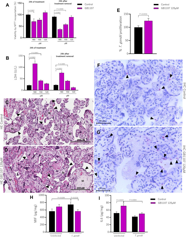
Galectin-3 (Gal-3) is a β-galactoside-binding lectin expressed in cells of the placental microenvironment. This lectin is involved in various biological processes, such as modulation of the immune system and control of parasitic illness. Toxoplasma gondii infection can lead to congenital transmission and cause miscarriages, prematurity and fetal anomalies. However, little is known about the role of Gal-3 in T. gondii infection in the placental microenvironment. This study aimed to unravel the underlying mechanisms of Gal-3 during T. gondii infection. For this purpose, we promoted the knockdown of Gal-3 expression by using RNA interference (RNAi) in BeWo cells or by using a synthetic inhibitor (GB1107) in human villous explants. We showed that the decreased Gal-3 expression in BeWo cells and human villous explants increases the invasion and proliferation of T. gondii probably by downregulating MIF and IL6 levels, highlighting thus the role of this lectin in modulating the immune response. Collectively, our study reveals Gal-3 as a promising target protein during congenital toxoplasmosis.

Keywords: Gal-3; congenital toxoplasmosis; immune response; maternal-fetal interface; placental.


Conflict of interest statement

The authors declare that the research was conducted in the absence of any commercial or financial relationships that could be construed as a potential conflict of interest.

Figures

Figure 1
Figure 1
Galectin-3 expression is efficiently reduced in BeWo cells after shRNA transfection. BeWo cells were transfected with Galectin-3 shRNA or control shRNA lentiviral particles. The knockdown of Gal-3 was confirmed by western blotting (A) and immunofluorescence assays (B–D). (E) Graphical representation of the mean of fluorescence intensity (MFI) of Gal-3 expression. Photomicrographs were captured at 40x magnification. The cell nucleus is labeled with TO-PRO-3 (blue); Gal-3 labeled with Alexa Fluor 488-conjugated is shown green. Scale bar 24µm.
Figure 2
Figure 2
The knockdown of Galectin-3 in BeWo cells increases the invasion and intracellular proliferation of T. gondii. (A) BeWo cells silenced or not for Gal-3 were infected for 3h to evaluate T. gondii invasion or (B) for 24h to evaluate parasite proliferation. In both experimental situations, the number of tachyzoites was determined by β-galactosidase assay and expressed as percentage. The proportion of the number of intracellular tachyzoites in relation to the total number of parasites was considered as the invasion ratio (C). The number of intracellular (green+/red) and attached [red or red+/green+ (yellow)] parasites was scored in 20 randomly selected fields and expressed as the ratio of the number of parasites per cell nucleus (D). Representative fluorescence images highlighting the impact of the decrease of Gal-3 expression on parasite proliferation (E–G). Photomicrographs were captured at 40x magnification (Scale bar: 24μm). Data are presented as means ± standard error of the means (SEM). Significant differences were analyzed using one-way ANOVA with Sidak’s multiple comparison post-test. Differences were considered statistically significant when P < 0.05.
Figure 3
Figure 3
The knockdown of Galectin-3 reduces the production of MIF and IL6 in T. gondii-infected BeWo cells. Wild Type or shGal-3 BeWo cells were infected or not with T. gondii tachyzoites and cell culture supernatants were collected for measurement of (A) MIF, (B) IL6, (C) IL8 and (D) IFNγ. (E) Infected cells were incubated with H2DCF-DA, and the ROS production was expressed as median fluorescence intensity of H2DCFDA. Data are shown as mean ± standard error of the means (SEM). Differences were analyzed using one-way ANOVA with Sidak’s multiple comparison post-test. Differences were considered statistically significant when P < 0.05.
Figure 4
Figure 4
Treatment with synthetic inhibitors (GB1107) does not affect the viability of placental villi and increases parasitism. Placental villi were incubated for 24h with GB1107 (180, 150, 120µM), and subsequently the treatment was removed and only culture medium was added for another 24h and subjected to viability analysis. (A) Viability of placental villi was determined by MTT incorporation, with the control group considered as 100% viable. (B) Supernatants from placental villi were collected and used to measure LDH levels (U/L). Representative photomicrographs of hematoxylin-eosin (HE) stained histological sections of placental villi treated with (C) culture medium or (D) inhibitor (120µM) for 24h are shown [syncytiotrophoblast cells (black arrowhead) and mesenchyme (M)]. Photomicrographs were captured at 40x magnification (scale bar: 200µm). (E) Placental villi previously incubated with inhibitor (120µM) were infected with T. gondii tachyzoites for 24h, and the percentage of intracellular parasite proliferation was measured by the β-galactosidase assay, with untreated/infected (control group) considered as 100% parasite proliferation. (F, G) Representative photomicrographs show T. gondii tachyzoites (black arrowhead) immunolocated by immunophosphatase staining and counterstained with hematoxylin (scale bar: 50μm). Additionally, placental villi were infected or not with T. gondii tachyzoites, and cell culture supernatants were collected for measurement of (H) MIF, (I) IL6. Data are shown as mean ± standard error of the means (SEM). Differences were analyzed using one-way ANOVA with Sidak’s multiple comparison post-test. Differences were considered statistically significant when P < 0.05.

References

    1. Aguirre A. A., Longcore T., Barbieri M., Dabritz H., Hill D., Klein P. N., et al. . (2019). The one health approach to toxoplasmosis: epidemiology, control, and prevention strategies. Ecohealth 16, 378–390. doi: 10.1007/s10393-019-01405-7 - DOI - PMC - PubMed
    1. Alves C. M., Silva D. A., Azzolini A. E., Marzocchi-MaChado C. M., Carvalho J. V., Pajuaba A. C., et al. . (2010). Galectin-3 plays a modulatory role in the life span and activation of murine neutrophils during early Toxoplasma gondii infection. Immunobiology 215, 475–485. doi: 10.1016/j.imbio.2009.08.001 - DOI - PubMed
    1. Alves C. M., Silva D. A., Azzolini A. E., Marzocchi-MaChado C. M., Lucisano-Valim Y. M., Roque-Barreira M. C., et al. . (2013). Galectin-3 is essential for reactive oxygen species production by peritoneal neutrophils from mice infected with a virulent strain of Toxoplasma gondii. Parasitology 140, 210–219. doi: 10.1017/s0031182012001473 - DOI - PubMed
    1. Barbosa B. F., Lopes-Maria J. B., Gomes A. O., Angeloni M. B., Castro A. S., Franco P. S., et al. . (2015). IL10, TGF beta1, and IFN gamma modulate intracellular signaling pathways and cytokine production to control Toxoplasma gondii infection in BeWo trophoblast cells. Biol. Reprod. 92, 82. doi: 10.1095/biolreprod.114.124115 - DOI - PubMed
    1. Bernardes E. S., Silva N. M., Ruas L. P., Mineo J. R., Loyola A. M., Hsu D. K., et al. . (2006). Toxoplasma gondii infection reveals a novel regulatory role for galectin-3 in the interface of innate and adaptive immunity. Am. J. Pathol. 168, 1910–1920. doi: 10.2353/ajpath.2006.050636 - DOI - PMC - PubMed

MeSH terms

LinkOut - more resources