

Case Reports
doi: 10.1016/j.eucr.2024.102882.
eCollection 2025 Jan.
Urethral clear cell adenocarcinoma in an adult female: A rare case report
Affiliations
- PMID: 39655199
- PMCID: PMC11626717
- DOI: 10.1016/j.eucr.2024.102882
Item in Clipboard
Case Reports
Urethral clear cell adenocarcinoma in an adult female: A rare case report
Urol Case Rep.
.
Abstract
Clear cell adenocarcinoma of the urethra is an extremely rare malignancy with a poor outcome, mainly affecting females in old age. We present the case of a 42-year-old female patient who presented with progressively worsening lower urinary tract symptoms, leading to a cystoscopy-guided core needle biopsy diagnosis of clear cell adenocarcinoma of the urethra. We will mainly discuss the cross-sectional imaging and pathological aspects of the case.
© 2024 The Authors.
Conflict of interest statement
The authors declare that they have no known competing financial interests or personal relationships that could have appeared to influence the work reported in this paper.
Figures
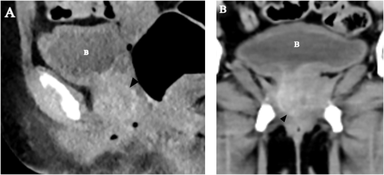
Axial (A) and coronal (B) post-contrast pelvic CT show bulky and predominantly homogeneously enhancing urethral lesion with bladder base extension (arrowhead in A and B).Bladder is labelled B in both images.

Sagittal MRI in T1W (A), T2W (B), and T2W fat-suppressed (C) sequences: The entire urethra shows a T1 intermediate as well as T2 and T2 FS hyperintense bulky tumoral infiltration with luminal obliteration. The normal zonal anatomy of the urethra is lost. The lesion has anterior vaginal (white arrowheads) and bladder (B) base extensions.

Post-contrast T1W Volumetric MRI: The tumor shows marked enhancement along its entire height and width. The bladder extension is better appreciated.

A and B- Hematoxylin and Eosin stain (400x) - Cuboidal cells have hyperchromatic nuclei, abundant clear to pale eosinophilic cytoplasm and hobnailing.

Immunohistochemical staining for PAX8, 400x (A) and CK7, 100x (B) - The tumor shows diffuse nuclear positivity for PAX8 and diffuse membranous positivity for CK7.
References
-
- Oliva E., Young R.H. Clear cell adenocarcinoma of the urethra: a clinicopathologic analysis of 19 cases. Mod Pathol. 1996 May;9(5):513–520. - PubMed
-
- Sheahan G., Vega A. Primary Clear Cell Adenocarcinoma in a Female Urethral Diverticulum: A Case Report and Review. World J Nephrol Urol. 2013;2(1):29–32.
-
- Sung M.T., Zhang S., MacLennan G.T., et al. Histogenesis of clear cell adenocarcinoma in the urinary tract: evidence of urothelial origin. Clin Cancer Res. 2008 Apr 1;14(7):1947–1955. - PubMed
-
- MRI of female urethral and periurethral disorders. Am J Rev. 2024 https://www.ajronline.org/doi/10.2214/ajr.182.3.1820677 - DOI - PubMed
Publication types
LinkOut - more resources
Full Text Sources
