Review
. 2024 Nov 25:18:1477753.
doi: 10.3389/fncel.2024.1477753. eCollection 2024.

Interlaminar and varicose-projection astrocytes: toward a new understanding of the primate brain

Affiliations
Review

Interlaminar and varicose-projection astrocytes: toward a new understanding of the primate brain

Caterina Ciani et al. Front Cell Neurosci. .

Abstract

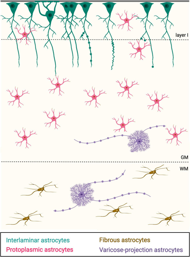
In the last years, science started to move toward a more glio-neurocentric view, in which astrocytes are hypothesized to be directly involved in cognitive functions. Indeed, astrocytes show a variety of shapes with species-specific characteristics, suggesting a specialization of roles during evolution. Interlaminar (ILA) and varicose-projection (VP-As) astrocytes show an anatomical organization that is different compared to the classical horizontal net typically formed by protoplasmic and fibrous astrocytes. ILAs show a modular architecture with the soma in the first cortical layer and processes toward the deep layers with species-specific length. VP-As reside in the deep layers of the cortex, are characterized by varicosities on the longest processes, and are individual-specific. These characteristics suggest roles that are more complex than what was theorized until now. Here, we recapitulate what we know so far from literature from the first time ILAs were described to the most recent discoveries, spanning from morphology description, hypothesis on the development to their features in diseases. For a complete glance on this topic, we included a final paragraph on which techniques and models were used to study ILAs and VP-As, and what new avenues may be opened thanks to more novel methods.

Keywords: varicose-projection astrocytes; astrocytes; cerebral cortex; evolution; interlaminar astrocytes; primates.


Conflict of interest statement

The authors declare that the research was conducted in the absence of any commercial or financial relationships that could be construed as a potential conflict of interest.

Figures

FIGURE 1
FIGURE 1
Schematic representation of the four main types of astrocytes recognizable based on their morphology. In teal, interlaminar astrocytes; in magenta: protoplasmic astrocytes; in brown, fibrous astrocytes; in violet, varicose-projection astrocytes. Pia is on top. GM, gray matter; WM, white matter.
FIGURE 2
FIGURE 2
Example of GFAP+ interlaminar astrocytes and their processes from the cerebral cortex of primates [Adapted from Falcone et al. (2019) and Reisin and Colombo (2002)). (A) Example of an ILA in the prefrontal cortex of rhesus macaque (Macaca mulatta), with pia on top of the image. Immunofluorescence staining: DAPI in blue, GFAP in green, Lectin in red. Scale bar = 20 μm. (B) Representative image of GFAP+ pial ILAs in bonobo (Pan paniscus) dorsal cortex, immuno-stained with DAB. Scale bar = 50 μm. (C–E) Arrowheads indicate the cytoskeletal terminal masses decorating the endings of ILA processes, immunostained with anti-GFAP antibody. Large arrows indicate the direction of the cortical surface. (C) Brodmann Area 10, age not available; (D) Brodmann Area 17/ 18, age 65; (E) Brodmann Area 4, age 44. Scale bar in (C) = 18 um; scale bar in (D,E) = 20 μm.
FIGURE 3
FIGURE 3
Evolutionary tree showing the phylogenetic relationships of the species mentioned in this review. This tree was created with https://phylot.biobyte.de, species taxonomy codes were obtained from NCBI taxonomy database. Color code indicates closely related mammalian orders or clades. It is as follows: magenta for Marsupials, gray for Proboscidea, light blue for Carnivora, dark blue for Artiodactyls, purple for Chiroptera, yellow for Scadentia, brown for Rodents, green for Primates (specifically, light green for Strepsirrhini, medium green for New and Old World monkeys, dark green for humans and apes).

References

    1. Aamodt S. (2007). Focus on glia and disease. Nat. Neurosci. 10:1349. - PubMed
    1. Amadio S., Montilli C., Magliozzi R., Bernardi G., Reynolds R., Volonté C. (2010). P2Y12 receptor protein in cortical gray matter lesions in multiple sclerosis. Cerebr. Cortex 20 1263–1273. 10.1093/cercor/bhp193 - DOI - PubMed
    1. Amin N., De Vrij F. M. S., Baghdadi M., Brouwer R. W. W., Van Rooij J. G. J., Jovanova O., et al. (2018). A rare missense variant in RCL1 segregates with depression in extended families. Mol. Psychiatry 23 1120–1126. 10.1038/mp.2017.49 - DOI - PMC - PubMed
    1. Andriezen W. (1893). The neuroglia elements in the human brain. Br. Med. J. 2 227–230. 10.1136/bmj.2.1700.227 - DOI - PMC - PubMed
    1. Besser L. (1866). Zur Histogenese der Nerviiscn Elementartheile in Centralorganen des Neugebornen Menschen. Berlin: Springer

LinkOut - more resources