. 2024 Nov 23:40:101884.
doi: 10.1016/j.bbrep.2024.101884. eCollection 2024 Dec.

Delphinidin induces a fast-to-slow muscle fiber type shift through the AMPK signaling pathway in C2C12 myotubes

Affiliations

Delphinidin induces a fast-to-slow muscle fiber type shift through the AMPK signaling pathway in C2C12 myotubes

Motoki Murata et al. Biochem Biophys Rep. .

Abstract

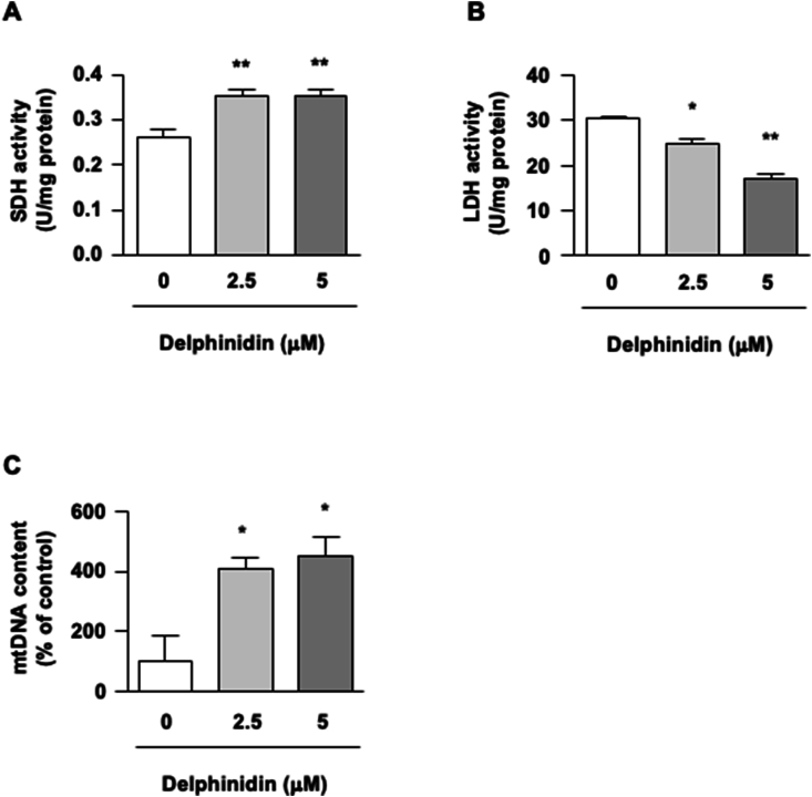
Delphinidin, a plant anthocyanidin, suppresses disuse muscle atrophy in mice. However, its effect on muscle fiber type shift is unclear. To examine whether delphinidin affects skeletal muscle fiber type, differentiated C2C12 cells were treated with delphinidin. Results revealed that delphinidin upregulated the mRNA expression of myosin heavy chain type I (MyHCI), troponin C1, troponin I1, and MyHCIIx and increased slow MyHC protein level in C2C12 myotubes. Delphinidin also enhanced succinic dehydrogenase (SDH) activities and suppressed lactate dehydrogenase (LDH) activity. Adenosine monophosphate-activated protein kinase (AMPK) inhibition attenuated delphinidin-induced MyHCI upregulation and MyHCIIb downregulation. We investigated the effect of delphinidin on the upstream factors involved in AMPK activation. Delphinidin increased liver kinase B1 (LKB1) phosphorylation and nuclear respiratory factor 1 (NRF1) and calcium/calmodulin-dependent protein kinase 2 (CaMKK2) protein levels. In conclusion, delphinidin induced muscle fiber type conversion from fast-twitch to slow-twitch muscles through the AMPK signaling pathway.

Keywords: AMPK; Anthocyanidin; C2C12 myotube; MyHC.


Conflict of interest statement

The authors declare that they have no known competing financial interests or personal relationships that could have appeared to influence the work reported in this paper.

Figures

Image 1
Graphical abstract
Fig. 1
Fig. 1
Delphinidin induces muscle fiber type shift in C2C12 myotubes. (A) Chemical structure of typical anthocyanidins. (B) C2C12 myotubes were treated with 5 μM anthocyanidin for 48 h. We conducted qRT-PCR to measure the mRNA expression of MyHCI and MyHCIIb. (C, D) C2C12 myotubes were treated with 2.5 μM or 5 μM delphinidin. After 48 h of treatment, the mRNA expression levels were measured via qRT-PCR. After 72 h of treatment, the protein levels of slow MyHC and fast MyHC were measured by western blotting. Data are presented as mean ± SEM, n = 3. ∗P < 0.05 and ∗∗P < 0.01 as compared with Control or 0 μM delphinidin.
Fig. 2
Fig. 2
Effect of delphinidin on metabolic enzyme activities and mtDNA content in C2C12 myotubes. (A, B) C2C12 myotubes were incubated with delphinidin for 72 h, and the enzyme activities were measured using commercial kits. (C) Mitochondrial DNA (mtDNA) in C2C12 cells treated with delphinidin for 72 h was analyzed by qRT-PCR. Data are presented as mean ± SEM, n = 3. ∗P < 0.05 and ∗∗P < 0.01 as compared with 0 μM delphinidin.
Fig. 3
Fig. 3
Delphinidin promotes muscle fiber type shift through AMPK in C2C12 myotubes. (A) C2C12 myotubes were treated with delphinidin for 1 h; subsequently, the phosphorylation of AMPK was assessed by western blotting and then normalized to AMPK. (B) After the C2C12 myotubes were treated with delphinidin for 48 h, Sirt1 and PGC-1α mRNA expression levels were measured by qRT-PCR. (C) C2C12 myotubes were treated with 5 μM delphinidin and 10 μM compound C separately or in combination for 48 h. Next, the mRNA expression levels of MyHCI and MyHCIIb were measured by qRT-PCR. (D) C2C12 myotubes were treated with 5 μM delphinidin and 10 μM compound C separately or in combination for 1 h. The protein level of p-AMPK was determined by western blotting analysis and normalized to AMPK. Data are presented as mean ± SEM, n = 3. ∗P < 0.05, ∗∗P < 0.01, and ∗∗∗P < 0.001 as compared with 0 μM delphinidin.
Fig. 4
Fig. 4
Delphinidin regulates the upstream the factors of AMPK signaling in C2C12 myotubes. (A, B) After the C2C12 myotubes were treated with delphinidin for 1 h, the protein level was assessed by western blotting. Data are presented as mean ± SEM, n = 3. ∗P < 0.05 as compared with 0 μM delphinidin.

References

    1. Alappat B., Alappat J. Anthocyanin pigments: beyond aesthetics. Molecules. 2020;25:5500. doi: 10.3390/molecules25235500. - DOI - PMC - PubMed
    1. Li D., Wang P., Luo Y., Zhao M., Chen F. Health benefits of anthocyanins and molecular mechanisms: update from recent decade. Crit. Rev. Food Sci. Nutr. 2017;57:1729–1741. doi: 10.1080/10408398.2015.1030064. - DOI - PubMed
    1. Huang C.C., Hung C.H., Hung T.W., Lin Y.C., Wang C.J., Kao S.H. Dietary delphinidin inhibits human colorectal cancer metastasis associating with upregulation of miR-204-3p and suppression of the integrin/FAK axis. Sci. Rep. 2019;9 doi: 10.1038/s41598-019-55505-z. - DOI - PMC - PubMed
    1. Sogo T., Terahara N., Hisanaga A., Kumamoto T., Yamashiro T., Wu S., Sakao K., Hou D.X. Anti-inflammatory activity and molecular mechanism of delphinidin 3-sambubioside, a Hibiscus anthocyanin. Biofactors. 2015;41:58–65. doi: 10.1002/biof.1201. - DOI - PubMed
    1. Park M., Sharma A., Lee H.J. Anti-adipogenic effects of delphinidin-3-O-β-glucoside in 3T3-L1 preadipocytes and primary white adipocytes. Molecules. 2019;24:1848. doi: 10.3390/molecules24101848. - DOI - PMC - PubMed

LinkOut - more resources