Case Reports
. 2025 Jan;21(1):e14394.
doi: 10.1002/alz.14394. Epub 2024 Dec 10.

Neuropathology of trisomy 21 mosaicism in a case with early-onset dementia

Affiliations
Case Reports

Neuropathology of trisomy 21 mosaicism in a case with early-onset dementia

Phong T Ngo et al. Alzheimers Dement. 2025 Jan.

Abstract

Introduction: This study investigated the impact of trisomy 21 mosaicism (mT21) on Alzheimer's disease (AD) neuropathology in a well-characterized clinical case described by Ringman et al.

Methods: We describe AD neuropathology in mT21 including amyloid beta, phosphorylated tau, astrogliosis, microgliosis, α-synuclein, and TAR DNA-binding protein 43 (TDP-43) in cerebral cortex, hippocampal subregions, and amygdala using immunohistochemistry.

Results: We observed high AD neuropathologic change with a score of A3B3C3. In addition, there was widespread astrogliosis, cerebral amyloid angiopathy, and perivascular space widening throughout the brain. Lewy bodies and neurites were noted in the amygdala only and no TDP-43 was observed.

Discussion: The findings in this case report highlight that mT21 is sufficient to induce AD neuropathology and early-onset dementia.

Highlights: Trisomy 21 mosaicism (mT21) occurs when three copies of chromosome 21 are present in some but not all somatic cells in an individual. mT21 accounts for ≈ 2% of people diagnosed with Down syndrome (DS). Immunohistochemical identification of amyloid beta, tau, astrocytes, microglia, α-synuclein, and TAR DNA-binding protein 43 show that Alzheimer's disease (AD) pathology in mT21 is similar to full trisomy 21. The findings in this case report highlight that mT21 is sufficient to induce AD neuropathology and early-onset dementia.

Keywords: Alzheimer's disease; Down syndrome; amyloid beta; neurofibrillary tangles; perivascular space widening.


Conflict of interest statement

The authors declare there are no competing interests. Author disclosures are available in the supporting information.

Figures
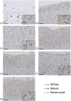
FIGURE 1
FIGURE 1
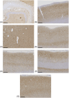
Amyloid beta (Aβ) plaques were observed throughout the individual with trisomy 21 mosaicism's brain. Low‐power images were taken with a 4 × objective lens with 500 µm scale bars. All inset images were taken using a 20 × objective lens with 100 µm scale bars. (A) Plaques in the hippocampus with an inset image of mature and diffuse plaques observed in the CA1. (B) The entorhinal cortex presented with dense‐cored, mature, and fibrillar plaques with neurons captured inside as seen in the inset image. Plaques in the (C) amygdala and (D)superior temporal lobe (E) parietal lobe are shown. (F) The occipital lobe contained small compact and cored plaques as well as dense fibrillar Aβ deposits. (G) In the dorsolateral prefrontal region, we observed many cored and compact plaques of smaller sizes.
FIGURE 2
FIGURE 2
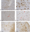
AT8 immunohistochemical staining for hyperphosphorylated tau proteins in the brain of the individual with trisomy 21 mosaicism. All images were taken using a 4 × objective lens with 500 µm scale bars. (A) In the hippocampus proper, large NPs were observed in the dentate gyrus region. NFTs and NTs were observed throughout the cornu ammonis sectors. Many NFTs and NTs were seen in the entorhinal cortex (B), amygdala (C), superior temporal lobe (D), and parietal lobe (E). Large, noticeable NPs were observed in the occipital lobe (F) and dorsolateral prefrontal region (G). NFTs, neurofibrillary tangles; NPs, neuritic plaques; NTs, neuropil threads.
FIGURE 3
FIGURE 3
Cerebral amyloid angiopathy and glial pathology in the individual with trisomy 21 mosaicism. (A) A 4 × image depicting the extensive presence of cerebral amyloid angiopathy within the dorsolateral prefrontal region in the form of amyloid beta (Aβ) deposits in both leptomeningeal and cortical vessels in the brain. (B) A 20 × image capturing Aβ deposits lining the entire cross‐sectional view of a cortical vessel in the gray matter of the dorsolateral prefrontal region. Additionally, the enlarged perivascular space can be seen surrounding the cortical vessel. (C) Ramified, ionized calcium‐binding adapter molecule 1–positive microglia in the gray matter of the dorsolateral prefrontal region. Image was captured using a 20 × objective lens. (D) As indicated by the red arrows, we observed rod‐shaped microglia in the cornu ammonis sector 3 (20 × image). (E) Hypertrophic, glial fibrillary acidic protein–positive astrocytes were seen interacting with blood vessels with enlarged perivascular spaces surrounding cortical vessels in the cornu ammonis sector 1 region of the hippocampus proper. The image was captured using a 10 × objective lens. (F)22 A 10 × image of hypertrophic astrocytes in the gray matter of the occipital lobe. The astrocytes can be seen in clusters with their processes surrounding spherical objects—presumably plaques. Scale bar = 500 µm (A), 200 µm (E–F), and 100 µm (B–D).

References

    1. Mai CT, Isenburg JL, Canfield MA, et al. National population‐based estimates for major birth defects, 2010‐2014. Birth Defects Res. 2019;111(18):1420‐1435. - PMC - PubMed
    1. Down JL. Observations on an ethnic classification of idiots. 1866. Ment Retard. 1995;33(1):54‐56. - PubMed
    1. Shin M, Siffel C, Correa A. Survival of children with mosaic Down syndrome. Am J Med Genet A. 2010;152A(3):800‐801. - PubMed
    1. Papavassiliou P, Charalsawadi C, Rafferty K, Jackson‐Cook C. Mosaicism for trisomy 21: a review. Am J Med Genet A. 2015;167A(1):26‐39. - PubMed
    1. Rumble B, Retallack R, Hilbich C, et al. Amyloid A4 protein and its precursor in Down's syndrome and Alzheimer's disease. N Engl J Med. 1989;320(22):1446‐1452. - PubMed

Publication types