. 2024 Dec;532(12):e70003.
doi: 10.1002/cne.70003.

Developmental and Adult Striatal Patterning of Nociceptin Ligand Marks Striosomal Population With Direct Dopamine Projections

Affiliations

Developmental and Adult Striatal Patterning of Nociceptin Ligand Marks Striosomal Population With Direct Dopamine Projections

Emily Hueske et al. J Comp Neurol. 2024 Dec.

Abstract

Circuit influences on the midbrain dopamine system are crucial to adaptive behavior and cognition. Recent developments in the study of neuropeptide systems have enabled high-resolution investigations of the intersection of neuromodulatory signals with basal ganglia circuitry, identifying the nociceptin/orphanin FQ (N/OFQ) endogenous opioid peptide system as a prospective regulator of striatal dopamine signaling. Using a prepronociceptin-Cre reporter mouse line, we characterized highly selective striosomal patterning of Pnoc mRNA expression in mouse dorsal striatum, reflecting the early developmental expression of Pnoc. In the ventral striatum, Pnoc expression in the nucleus accumbens core was grouped in clusters akin to the distribution found in striosomes. We found that PnoctdTomato reporter cells largely comprise a population of dopamine receptor D1 (Drd1) expressing medium spiny projection neurons localized in dorsal striosomes, known to be unique among striatal projection neurons for their direct innervation of midbrain dopamine neurons. These findings provide a new understanding of the intersection of the N/OFQ system among basal ganglia circuits with particular implications for developmental regulation or wiring of striato-nigral circuits.

Keywords: dendron bouquet; dopamine; nociceptin; opioid; orphanin F/Q; striatum; striosome.


Figures

FIGURE 1
FIGURE 1
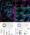
PnoctdTomato reporter expression shows compartmental distribution across the striatum and corresponds with striosomes in the dorsal striatum. (a) Composite image showing PnoctdTomato (magenta) and striosomal marker MOR1 (cyan) in the striatum. (b–d) Higher magnification images of the top boxed region from panel (a) with PnoctdTomato (b, magenta) and MOR1 (c, cyan) shown separately. Merged image (d) shows strong overlap between PnoctdTomato neurons and MOR1 expression. (e–g) same as (b–d) but for the lower boxed region in the ventral striatum from panel (a). (h) Quantification of PnoctdTomato spatial distribution across the dorsal and ventral (nucleus accumbens core, medial shell, and lateral shell) striatum (bar graph, one‐way ANOVA with Bonferroni's multiple comparisons test, ****p < 0.0001, N = 12 mice, N = 19 sections, 1–2 sections/mouse. Data represented as mean ± SEM across mice). (i) Quantification of PnoctdTomato spatial localization in striosomes vs. the matrix across the anterior to posterior axis in the dorsal striatum (two‐way ANOVA shows main effect of striatal compartment ***p < 0.001, no significant effect of anterior‐posterior axis p = 0.0851 bar graph, two‐way ANOVA with Bonferroni's multiple comparisons test, ***p < 0.001, ****p < 0.0001, N = 8 mice, 3 sections/mouse). Data represented as mean ± SEM across mice. ANT = anterior; DS = dorsal striatum; lNAcSh = lateral accumbens shell; MID = medial; mNAcSh = medial accumbens shell; NAc = nucleus accumbens; NAcC = nucleus accumbens core; POS = posterior.
FIGURE 2
FIGURE 2
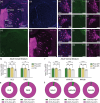
PnoctdTomato expressing striatal neurons cluster with Drd1 SPNs to near exclusion of Drd2. (a) Sagittal image of PnoctdTomato cells (magenta) colocalizing with D1‐GFP‐expressing SPNs (green) in dorsal (b–e) and ventral (f–i) striatum. (b) Higher magnification image of top boxed region from panel (a) in the dorsal striatum. (c–e) Higher magnification images from inset in (b) of dorsal striatum. Arrowheads point to cells expressing markers for both PnoctdTomato and D1‐GFP. (f) Higher magnification image of the bottom boxed region from panel (a) in the ventral striatum (nucleus accumbens). (g–i) Higher magnification images from inset in (f) of the ventral striatum. (j) Sagittal image of minimal overlap between PnoctdTomato cells (magenta) and D2‐GFP expression (green) in dorsal (k–n) and ventral (o–r) striatum (same as [b–e] and [f–i] but for D2‐GFP expression). (s) Summary of IHC‐stained PnoctdTomato reporter cell colocalization with in situ hybridization‐labeled Drd1 and Drd2 SPNs in dorsal striatum. (t) Quantification of PnoctdTomato cells on Drd1 and Drd2 labeled SPNs across the anterior to posterior axis in the dorsal striatum (main effect of neuron type, two‐way ANOVA, ****p < 0.0001; bar graph, two‐way ANOVA with Bonferroni's multiple comparisons test, ****p < 0.0001, N = 6 mice, 3 brain sections/mouse). (u) Same as (s) for the nucleus accumbens. (v) Quantification in nucleus accumbens of PnoctdTomato labeling in Drd1 and Drd2 SPNs (bar graph, one‐way ANOVA with Bonferroni's multiple comparisons test, ***p < 0.001, *p < 0.05, N = 6 mice, N = 8 sections, 1–2 sections/mouse). Data represented as mean ± SEM across mice. ANT = anterior; DS = dorsal striatum; MID = medial; NAc = nucleus accumbens; POS = posterior.
FIGURE 3
FIGURE 3
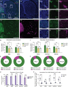
PnoctdTomato reporter expression marks a population of NPY and SST co‐expressing interneurons. (a) Coronal image of PnoctdTomato cells (magenta), somatostatin (white, SST), and neuropeptide Y (cyan, NPY) expression in the striatum. Higher magnification images of inset from panel (a) for (b) merged and (c–e) individual channels showing PnoctdTomato (c) colocalization with a subset of striatal interneurons co‐expressing SST (d) and NPY (e). (f–j) Same organization as (a–e) for additional interneuron markers; PnoctdTomato cells (magenta) show little colocalization with choline acetyltransferase (green, ChAT) or parvalbumin (blue, PV) expressing striatal interneurons. (k) Top: Summary of quantified PnoctdTomato cells colocalized with different interneuron markers in the dorsal striatum. Bottom: Quantification of PnoctdTomato cells colocalized with different interneuron markers across the anterior to posterior axis in the dorsal striatum (main effect of neuron type, two‐way ANOVA, ****p < 0.0001; bar graph, two‐way ANOVA with Bonferroni's multiple comparisons test, ****p < 0.0001, N = 6 mice, 3 sections/mouse). (l) Same as (k) but for nucleus accumbens (bar graph, one‐way ANOVA with Bonferroni's multiple comparisons test, ****p < 0.0001, **p < 0.01, *p < 0.05, N = 6 mice, N = 11 sections, 1–2 sections/mouse). Data represented as mean ± SEM across mice. ANT = anterior; lNAcSh = lateral accumbens shell; MID = medial; mNAcSh = medial accumbens shell; NAcC = nucleus accumbens core; POS = posterior.
FIGURE 4
FIGURE 4
Adult expression of striatal Pnoc mRNA is sparse relative to reporter‐labeled PnoctdTomato cells. (a–d) Coronal image showing composite (a) of DAPI (blue, b), Pnoc mRNA expression evaluated by ISH (green, c), and reporter‐labeled PnoctdTomato cells (magenta, d) in the adult striatum. Magnified insets from the dorsolateral striatum (e–g), dorsomedial striatum (h–j), nucleus accumbens core (k–m), and nucleus accumbens shell (n–p) show PnoctdTomato reporter‐labeled cells that do not co‐label with adult Pnoc mRNA expression. (q) Quantification of mean Pnoc mRNA intensity in PnoctdTomato cells (tdT+, green), nearby non‐reporter labeled cells (tdT, yellow), and in PnoctdTomato cells from adjacent control sections labeled with a negative control probe (NC, gray) in adult dorsal striatum. (Two‐way ANOVA with Bonferroni's multiple comparisons test, ***p < 0.001, **p < 0.01, ns = non‐significant, N = 4 mice, N = 7 sections, 1–2 sections/mouse). (r) Same as (q) but for adult ventral striatum (two‐way ANOVA with Bonferroni's multiple comparisons test, ****p < 0.0001, ***p < 0.001, ns = non‐significant, N = 4 mice, N = 7 sections, 1–2 sections/mouse). (s) Proportion of PnoctdTomato reporter cells identified as Pnoc mRNA positive (Pnoc+/tdT+, green) or negative (Pnoc/tdT+, magenta) in adult dorsal striatum. (t) Same as (s) but for adult ventral striatum. All data represented as mean ± SEM across mice. DLS = dorsolateral striatum; DMS = dorsomedial striatum; lNAcSh = lateral accumbens shell; mNAcSh = medial accumbens shell; NAcC = nucleus accumbens core.
FIGURE 5
FIGURE 5
Neonatal expression of striatal Pnoc mRNA recapitulates reporter‐labeled patterning in PnoctdTomato cells. (a–d) Coronal image showing composite (a) of DAPI (blue, b), Pnoc mRNA expression evaluated by ISH (green, c), and reporter‐labeled PnoctdTomato cells (magenta, d) in postnatal Day 1 (P1) brain sections. Magnified insets from the dorsolateral striatum (e–g), dorsomedial striatum (h–j), nucleus accumbens core (k–m), and nucleus accumbens shell (n–p) show PnoctdTomato reporter‐labeled cells co‐labeled with neonatal Pnoc mRNA expression. (q) Quantification of mean Pnoc mRNA intensity in PnoctdTomato cells (Pnoc/tdT+, green), nearby non‐reporter labeled cells (Pnoc/tdT, yellow), and in PnoctdTomato cells from adjacent control sections labeled with a negative control probe (NC, gray) in neonatal dorsal striatum. (Two‐way ANOVA with Bonferroni's multiple comparisons test, **p < 0.01, ns = non‐significant, N = 4 mice, N = 8 sections, 2 sections/mouse). (r) Same as (q) but for neonatal ventral striatum (two‐way ANOVA with Bonferroni's multiple comparisons test, ****p < 0.0001, ***p < 0.001, ns = non‐significant, N = 4 mice, N = 7 sections, 1–2 sections/mouse). (s) Proportion of PnoctdTomato reporter cells identified as Pnoc mRNA positive (Pnoc+/tdT+, green) or negative (Pnoc/tdT+, magenta) in neonatal dorsal striatum. (t) Same as (s) but for neonatal ventral striatum. (u) Proportion of PnoctdTomato reporter cells identified as Pnoc mRNA positive in neonate (light purple) vs. adult (dark purple) tissue (two‐way ANOVA with Bonferroni's multiple comparisons test, ****p < 0.0001, N = 4 mice/group, N = 7 sections, 1–2 sections/mouse). (v) Fold‐change in mean Pnoc mRNA intensity in tdT+ cells relative to background (tdT cells) in neonate (light purple) vs. adult (dark purple) tissue (two‐way ANOVA with Bonferroni's multiple comparisons test, ****p < 0.0001, ***p < 0.001, **p < 0.01, ns = non‐significant, N = 4 mice/group, N = 7 sections, 1–2 sections/mouse). All data represented as mean ± SEM across mice. DLS = dorsolateral striatum; DMS = dorsomedial striatum; NAcC = nucleus accumbens core; NAcSh = nucleus accumbens shell.
FIGURE 6
FIGURE 6
Striosomal fibers from PnoctdTomato targeted neurons project to the striosome‐dendron bouquet in SNc in both adults and neonates. In PnoctdTomato reporter mice (a), tdTomato‐labeled fibers (magenta) project to TH‐labeled dopamine neurons (green) in adults (b–d) and neonates (e–g). (h) Schematic of mouse genetic strategy by viral labeling of striosomal cells in Pnoc‐Cre;loxP‐Stop‐loxP‐FlpO mice with a Flp‐dependent AAV expressing membrane‐GFP (mGFP). Virally targeted mGFP‐expressing PnocFlp neurons (i–j) exhibit projections to dopamine dendrons in SNc (k–m).

Update of

References

    1. Al‐Hasani, R. , McCall J. G., Shin G., et al. 2015. “Distinct Subpopulations of Nucleus Accumbens Dynorphin Neurons Drive Aversion and Reward.” Neuron 87, no. 5: 1063–1077. - PMC - PubMed
    1. Allen, R. G. , Peng B., Pellegrino M. J., et al. 2001. “Altered Processing of Pro‐Orphanin FQ/Nociceptin and Pro‐Opiomelanocortin‐Derived Peptides in the Brains of Mice Expressing Defective Prohormone Convertase 2.” Journal of Neuroscience 21, no. 16: 5864–5870. - PMC - PubMed
    1. Amemori, K.‐I. , Amemori S., Gibson D. J., and Graybiel A. M.. 2018. “Striatal Microstimulation Induces Persistent and Repetitive Negative Decision‐Making Predicted by Striatal Beta‐Band Oscillation.” Neuron 99, no. 4: 829–841.e6. - PMC - PubMed
    1. Amemori, S. , Amemori K.‐I., Yoshida T., et al. 2020. “Microstimulation of Primate Neocortex Targeting Striosomes Induces Negative Decision‐Making.” European Journal of Neuroscience 51, no. 3: 731–741. - PMC - PubMed
    1. Amemori, K.‐I. , and Graybiel A. M.. 2012. “Localized Microstimulation of Primate Pregenual Cingulate Cortex Induces Negative Decision‐Making.” Nature Neuroscience 15, no. 5: 776–785. - PMC - PubMed

LinkOut - more resources