. 2024 Dec;28(23):e70274.
doi: 10.1111/jcmm.70274.

S-Nitrosylation of NOTCH1 Regulates Mesenchymal Stem Cells Differentiation Into Hepatocyte-Like Cells by Inhibiting Notch Signalling Pathway

Affiliations

S-Nitrosylation of NOTCH1 Regulates Mesenchymal Stem Cells Differentiation Into Hepatocyte-Like Cells by Inhibiting Notch Signalling Pathway

Xuesong Wang et al. J Cell Mol Med. 2024 Dec.

Abstract

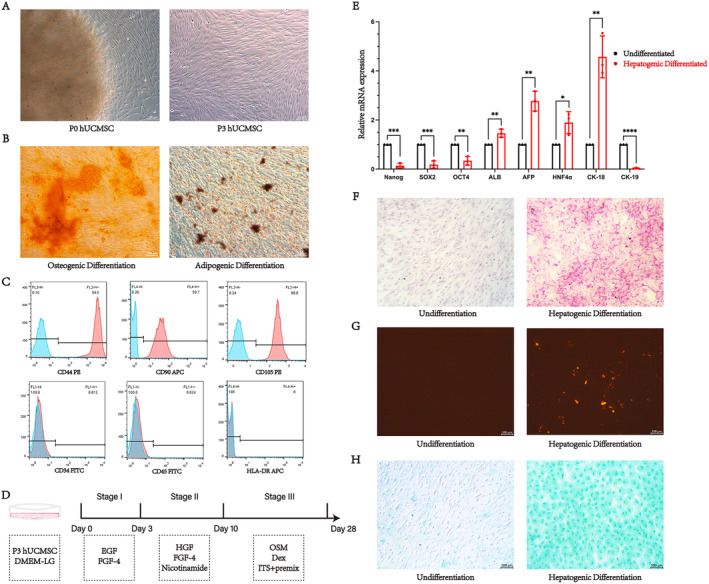
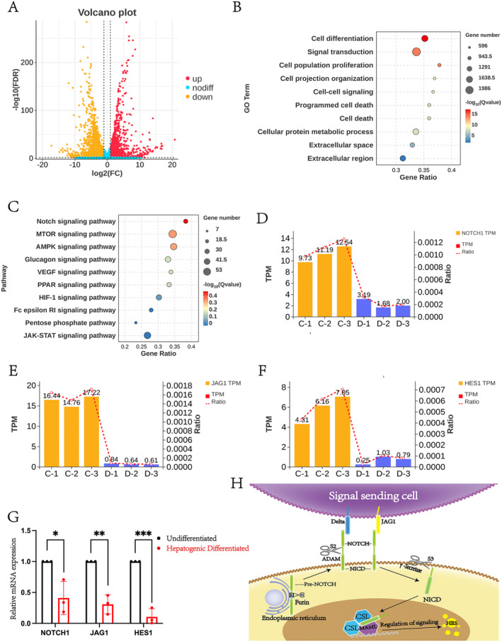
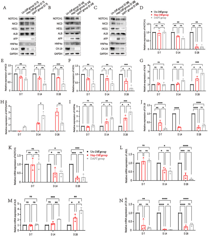
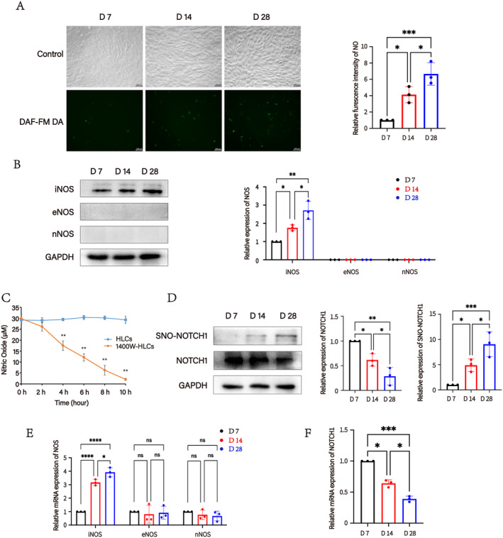
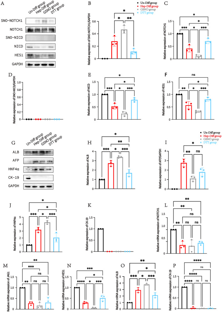
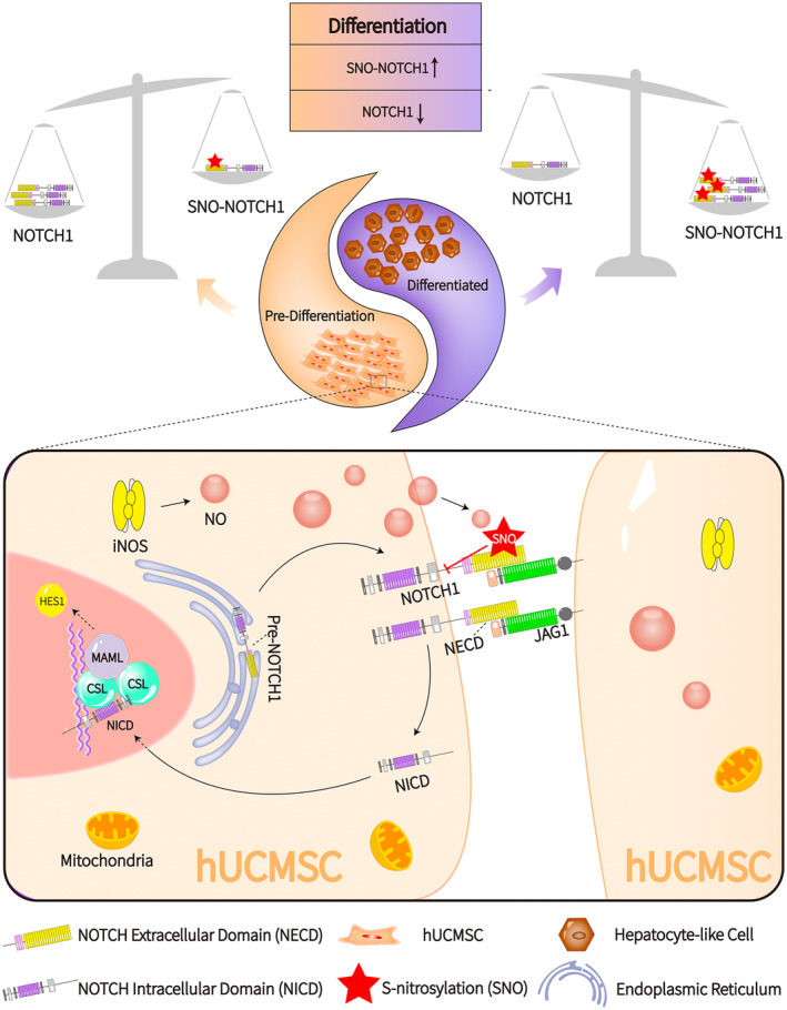
The differentiation of mesenchymal stem cells (MSCs) into hepatocyte-like cells (HLCs) is considered one of the most promising strategies for alternative hepatocyte transplantation to treat end-stage liver disease. To advance this method, it is crucial to gain a deeper understanding of the mechanisms governing hepatogenic differentiation. The study demonstrated that suppression of the intracellular domain release of the Notch pathway receptor via the γ-secretase inhibitor N-[(3, 5-difluorophenyl)acetyl]-L-alanyl-2-phenylglycine-1, 1-dimethylethyl ester (DAPT) significantly promotes the expression of hepatocyte-related genes and proteins in HLCs. Increased expression of intracellular inducible NO synthase (iNOS) during differentiation led to elevated endogenous NO production. Biotin switch assays revealed a gradual increase in S-nitrosylation (SNO)-NOTCH1 and a decrease in overall NOTCH1 expression during hepatogenic differentiation. The addition of the exogenous NO donor S-nitrosoglutathione (GSNO) and the SNO inhibitor dithiothreitol (DTT) further demonstrated that the elevated expression of SNO-NOTCH1 promotes the differentiation of MSCs into mature hepatocytes. Briefly, our results fully demonstrated that the modification of the extracellular domain of NOTCH1 by NO, leading to the formation of SNO-NOTCH1, significantly promotes hepatogenic differentiation by inhibiting the Notch signalling pathway. Our study highlights the critical role of SNO-NOTCH1 in regulating the Notch signalling pathway and offers new insights into the mechanisms driving this differentiation process.

Keywords: NO; S‐nitrosylation; differentiation mechanisms; hepatogenic differentiation; mesenchymal stem cells; signalling pathways.


Conflict of interest statement

The authors declare no conflicts of interest.

Figures

FIGURE 1
FIGURE 1
The characteristics of MSCs and their differentiation into functional HLCs in vitro. (A). Morphological characteristics of MSCs at P0 and P3. Scale bar = 100 μm. (B) Demonstration of the osteogenic and adipogenic differentiation potential of MSCs by Alizarin red staining and Oil Red O staining. Scale bar = 100 μm. (C) Expression of cell surface markers on MSCs. Cells were incubated with FITC, PE or APC‐conjugated anti‐human‐specific antibodies. Flow cytometry analysis revealed positive expression of CD44, CD90 and CD105, while CD34, CD45 and HLA‐DR were negative. (D) Induction protocol for differentiation of MSCs into HLCs. EGF. Epidermal growth factor; FGF4, Basic fibroblast growth factor; HGF, Hepatocyte growth factor; OSM, Oncostatin M; Dex, Dexamethasone. (E) RT‐qPCR analysis of mRNA expression levels of pluripotent transcription factors (NANOG, SOX2 and OCT4) and hepatocyte‐associated factors (ALB, AFP, HNF4α, CK‐18 and CK‐19) in the Un‐Diff group and the Hep‐Diff group. *P < 0.05, **P < 0.01, ***P < 0.001. (F) PAS staining demonstrating glycogen synthesis in Un‐Diff group and Hep‐Diff group. Scale bar = 100 μm. (G) Dil‐LDL labelling showing LDL uptake capacity of cells in Un‐Diff group and Hep‐Diff group. Scale bar = 100 μm. (H) ICG reagent demonstrating ICG uptake capacity of cells in Un‐Diff group and Hep‐Diff group. Scale bar, 100 μm.
FIGURE 2
FIGURE 2
The RNA‐seq results confirmed that the Notch signalling pathway is involved in the hepatogenic differentiation process of MSCs. (A) Volcano plot showing DEGs between the Un‐Diff group and the Hep‐Diff group. (B) Bubble plot of GO analysis for both the Un‐Diff group and the Hep‐Diff group. (C) KEGG analysis reveals that the Notch signalling pathways is associated with cellular signal transduction functions. (D) Expression levels of the Notch signalling pathway receptor NOTCH1 in both the Un‐Diff group and the Hep‐Diff group. C, Un‐Diff group; D, Hep‐Diff group; TPM, transcripts per million reads; Ratio, ratio of genes to total differential genes. (E) Expression levels of the Notch signalling pathway ligand JAG1 in both the Un‐Diff group and the Hep‐Diff group. C, Un‐Diff group; D, Hep‐Diff group; TPM, Transcripts per million reads; Ratio, Ratio of genes to total differential genes. (F) Expression levels of the Notch signalling pathway downstream factor HES1 in both the Un‐Diff group and the Hep‐Diff group. C, Un‐Diff group; D, Hep‐Diff group; TPM, transcripts per million reads; Ratio, ratio of genes to total differential genes. (G) RT‐qPCR detection of the relative mRNA expression levels of Notch signalling pathway factors (NOTCH1, JAG1 and HES1) in both the Un‐Diff group and the Hep‐Diff group. *p < 0.05, **p < 0.01, ***p < 0.001. (H) Diagram of the Notch signalling pathway.
FIGURE 3
FIGURE 3
The Notch signalling pathway regulates the differentiation of MSCs into HLCs. Western blotting and RT‐qPCR were employed to assess the expression differences of various factors in the Un‐Diff, Hep‐Diff and DAPT groups. (A) Protein expression of Notch signalling pathway factors (NOTCH1, NICD and HES1) and hepatocyte‐associated factors (ALB, AFP, HNF4α and CK‐19) was detected by Western blotting in each group at 7 days (D 7). (B) Protein expression of Notch signalling pathway factors (NOTCH1, NICD and HES1) and hepatocyte‐associated factors (ALB, AFP, HNF4α and CK‐19) was detected by Western blotting in each group at 14 days (D 14). (C) Protein expression of Notch signalling pathway factors (NOTCH1, NICD and HES1) and hepatocyte‐associated factors (ALB, AFP, HNF4α and CK‐19) was detected by Western blotting in each group at 28 days (D 28). (D) Changes in the relative protein expression of NOTCH1 in each group at differentiation time points of D 7, D 14 and D 28. **p < 0.01. (E) Changes in the relative protein expression of NICD in each group at differentiation time points of D 7, D 14 and D 28. *p < 0.05, **p < 0.01, ***p < 0.001. (F) Changes in the relative protein expression of HES1 in each group at differentiation time points at D 7, D 14 and D 28. *p < 0.05, **p < 0.01, ***p < 0.001. (G) Changes in the relative protein expression of ALB in each group at differentiation time points of D 7, D 14 and D 28. *p < 0.05, **p < 0.01, ***p < 0.001. (H) Changes in the relative protein expression of AFP in each group at differentiation time points of D 7, D 14 and D 28. *p < 0.05, **p < 0.01, ***p < 0.001. (I) Changes in the relative protein expression of HNF4α in each group at differentiation time points of D 7, D 14 and D 28. *p < 0.05, **p < 0.01, ***p < 0.001. (J) Changes in the relative protein expression of CK‐19 in each group at differentiation time points of D 7, D 14 and D 28. *p < 0.05, ***p < 0.001, ****p < 0.0001. (K) Changes in the relative mRNA expression of NOTCH1 in each group at differentiation time points of D 7, D 14 and D 28. *p < 0.05, ***p < 0.001. (L) Changes in the relative mRNA expression of JAG1 in each group at differentiation time points of D 7, D 14 and D 28. *p < 0.05, ***p < 0.001, ****p < 0.0001. (M) Changes in the relative mRNA expression of ALB in each group at differentiation time points of D 7, D 14 and D 28. *p < 0.05, **p < 0.001, ***p < 0.001. (N) Changes in the relative mRNA expression of CK‐19 in each group at differentiation time points of D 7, D 14 and D 28. *p < 0.05, **p < 0.01, ****p < 0.0001.
FIGURE 4
FIGURE 4
SNO‐NOTCH1 levels gradually increased while NOTCH1 protein levels gradually decreased during hepatogenic differentiation. (A) The fluorogram and relative expression of intracellular NO during hepatogenic differentiation for 7 days (D 7), 14 days (D 14) and 28 days (D 28). Scale bar = 100 μm. *p < 0.05, ***p < 0.001. (B) Relative protein expression of iNOS, eNOS and nNOS in cells undergoing hepatogenic differentiation for D 7, D 14 and D 28. *p < 0.05, **p < 0.01. (C) Effect of the selective iNOS inhibitor 1400 W on NO concentration in HLCs from 0 to 10 h. **p < 0.01. (D) Relative protein expression of NOTCH1 and relative expression of SNO‐NOTCH1 in MSCs undergoing hepatogenic differentiation for D 7, D 14 and D 28. *p < 0.05, **p < 0.01, ***p < 0.001. (E) Relative mRNA expression of iNOS, eNOS and nNOS in MSCs undergoing hepatogenic differentiation for D 7, D 14 and D 28. *p < 0.05, ****p < 0.0001. (F) Relative mRNA expression of NOTCH1 in MSCs undergoing hepatogenic differentiation for D 7, D 14 and D 28. *p < 0.05, ***p < 0.001.
FIGURE 5
FIGURE 5
S‐nitrosylation of NOTCH1 promotes Notch signalling pathway‐mediated hepatogenic differentiation by inhibiting NOTCH1 self‐expression. (A) Hepatogenic differentiation was observed for up to 28 days. This includes protein expression levels of NOTCH signalling pathway factors (NOTCH1, NICD and HES1) and S‐nitrosylation levels of NOTCH1 and NICD in the Un‐Diff, Hep‐Diff, GSNO and DTT groups. (B) Differences in relative protein expression of SNO‐NOTCH1 among the four groups. *p < 0.05, **p < 0.01. (C) Differences in relative protein expression of NOTCH1 among the four groups. *p < 0.05, ***p < 0.001. (D) Differences in relative protein expression of SNO‐NICD among the four groups. *p < 0.05, ***p < 0.001. (E) Differences in relative protein expression of NICD among the four groups. (F) Differences in relative protein expression of HES1 among the four groups. *p < 0.05, **p < 0.01, ***p < 0.001. (G) Hepatogenic differentiation was assessed up to 28 days. This includes protein expression levels of hepatocyte‐associated factors (ALB, AFP, CK‐18 and CK‐19) in the Un‐Diff, Hep‐Diff, GSNO, and DTT groups. (H) Differences in relative protein expression of ALB among the four groups. *p < 0.05, **p < 0.01, ***p < 0.001. (I) Differences in relative protein expression of AFP among the four groups. *p < 0.05, **p < 0.01. (J) Differences in relative protein expression of HNF4α among the four groups. *p < 0.05, ***p < 0.001. (K) Differences in relative protein expression of CK‐19 among the four groups. (L) Differences in relative mRNA expression of NOTCH1 among the four groups. **p < 0.01. (M) Differences in relative mRNA expression of JAG1 among the four groups. **p < 0.01, ***p < 0.001. (N) Differences in relative mRNA expression of HES1 among the four groups. *p < 0.05, **p < 0.01, ***p < 0.001. (O) Differences in relative mRNA expression of ALB among the four groups. *p < 0.05, **p < 0.01, ***p < 0.001. (P) Differences in relative mRNA expression of CK‐19 among the four groups. ****p < 0.0001.
FIGURE 6
FIGURE 6
The schematic diagram illustrates that the increase in NO content during the differentiation of MSCs into HLCs is due to the high expression of iNOS and that NO causes S‐nitrosative modification of the extracellular domain of NECD, which leads to the reduction in the expression level of NOTCH1, thereby inhibiting the Notch signalling pathway. Inhibition of the Notch signalling pathway promotes the process of hepatogenic differentiation. During the whole differentiation process, due to the presence of NO, SNO‐NOTCH1 and NOTCH1 are like in a Yin and Yang relationship, but closely linked.

Similar articles

References

    1. Budi N. Y. P., Lai W. Y., Huang Y. H., and Ho H. N., “3D Organoid Cultivation Improves the Maturation and Functional Differentiation of Cholangiocytes From Human Pluripotent Stem Cells,” Frontiers in Cell and Development Biology 12 (2024): 1361084. - PMC - PubMed
    1. Okumura A., Aoshima K., and Tanimizu N., “Generation of In Vivo‐Like Multicellular Liver Organoids by Mimicking Developmental Processes: A Review,” Regenerative Therapy 26 (2024): 219–234. - PMC - PubMed
    1. Arjunan P., Kathirvelu D., Mahalingam G., et al., “Lipid‐Nanoparticle‐Enabled Nucleic Acid Therapeutics for Liver Disorders,” Acta Pharmaceutica Sinica B 14, no. 7 (2024): 2885–2900. - PMC - PubMed
    1. Zeng Y., Wu Z., Chen G., et al., “Peripheral Injection of hUC‐MSCs in the Treatment of Acute Liver Failure: A Pre‐Clinical Cohort Study in Rhesus Monkeys,” Stem Cells International 2024 (2024): 4654912. - PMC - PubMed
    1. Wang H., Tian Y., Li X., Yang M., and Yan Y., “Amniotic Mesenchymal Stem Cells Derived Hepatocyte‐Like Cells Attenuated Liver Fibrosis More Efficiently by Mixed‐Cell Transplantation,” International Journal of Physiology, Pathophysiology and Pharmacology 12, no. 1 (2020): 11–24. - PMC - PubMed

MeSH terms

LinkOut - more resources