

The Organ-Joint Axes in Osteoarthritis: Significant Pathogenesis and Therapeutic Targets
- PMID: 39656496
- PMCID: PMC12339117
- DOI: 10.14336/AD.2024.1223
The Organ-Joint Axes in Osteoarthritis: Significant Pathogenesis and Therapeutic Targets
Abstract
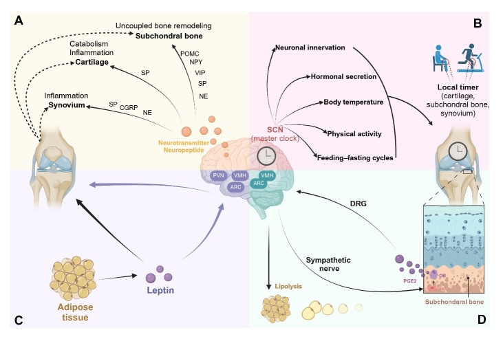
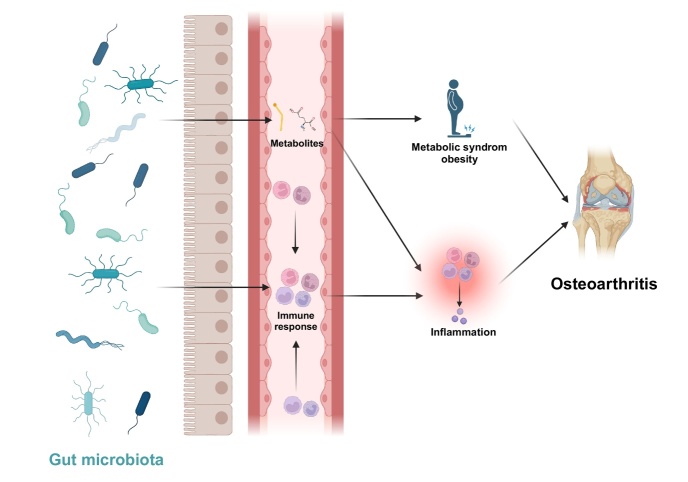
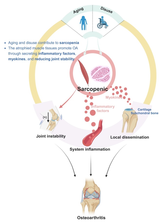
Osteoarthritis (OA), a prevalent age-related disease, is increasingly recognized as a multifactorial condition. This comprehensive review provides a multifaceted perspective on the organ-joint crosstalk contributing to OA, transcending the traditional focus on local joint pathology. Based on current research, we discussed the brain-joint, gut-joint, muscle-joint interactions in the etiology and progression of OA. In brain-joint axis, the neuroendocrine regulation, circadian rhythms, and leptin signaling influence joint tissues. We also discussed the role of prostaglandin E2 in skeletal interoception and its potential as a therapeutic target. The gut-joint axis is underscored by the impact of gut microbiota dysbiosis on systemic inflammation and metabolic disorders, both of which are implicated in OA pathogenesis. Furthermore, age-related sarcopenia, characterized by muscle mass and strength loss, is identified as a significant risk factor. Sarcopenia may contribute to OA progression through compromised mechanical support, systemic inflammation, and muscle-derived myokines. Finally, we synthesize the evidence supporting the modulation of circadian rhythm, skeletal interoception, gut microbiome, and muscle mass as innovative strategies for OA management. The organ-joint crosstalk is integral to the complex pathogenesis of OA, highlighting the multifactorial nature of OA and the potential for targeted therapeutic interventions. By integrating these multidimensional perspectives, we aim to enhance our understanding of OA pathogenesis and explore potential pharmacological targets.
Conflict of interest statement
The authors declare no competing interests.
Figures
Similar articles
-
Gut microbiota and osteoarthritis: epidemiology, mechanistic analysis, and new horizons for pharmacological interventions.Front Cell Infect Microbiol. 2025 Jul 16;15:1605860. doi: 10.3389/fcimb.2025.1605860. eCollection 2025. Front Cell Infect Microbiol. 2025. PMID: 40740348 Free PMC article. Review.
-
Gut microbiome dysbiosis accelerates osteoarthritis progression by inducing IFP-SM inflammation in "double-hit" mice.Arthritis Res Ther. 2025 Jul 7;27(1):137. doi: 10.1186/s13075-025-03602-y. Arthritis Res Ther. 2025. PMID: 40624668 Free PMC article.
-
Relationship Between the Gut Microbiome, Tryptophan-Derived Metabolites, and Osteoarthritis-Related Pain: A Systematic Review with Meta-Analysis.Nutrients. 2025 Jan 12;17(2):264. doi: 10.3390/nu17020264. Nutrients. 2025. PMID: 39861394 Free PMC article.
-
Osteoarthritis in cats: what we know, and mostly, what we don't know. . . yet.J Feline Med Surg. 2025 Jul;27(7):1098612X251347999. doi: 10.1177/1098612X251347999. Epub 2025 Jul 20. J Feline Med Surg. 2025. PMID: 40685570 Free PMC article. Review.
-
Exercise for hand osteoarthritis.Cochrane Database Syst Rev. 2017 Jan 31;1(1):CD010388. doi: 10.1002/14651858.CD010388.pub2. Cochrane Database Syst Rev. 2017. PMID: 28141914 Free PMC article.
Cited by
-
Gut microbiota and osteoarthritis: epidemiology, mechanistic analysis, and new horizons for pharmacological interventions.Front Cell Infect Microbiol. 2025 Jul 16;15:1605860. doi: 10.3389/fcimb.2025.1605860. eCollection 2025. Front Cell Infect Microbiol. 2025. PMID: 40740348 Free PMC article. Review.
References
-
- Lou Q, Jiang K, Wang X, Pan Y, Qiu G, Wu B, et al. (2024). IGF1R signaling in perinatal mesenchymal stem cells determines definitive hematopoiesis in bone marrow. Blood. - PubMed
-
- Lu K, Shi TS, Shen SY, Shi Y, Gao HL, Wu J, et al. (2022). Defects in a liver-bone axis contribute to hepatic osteodystrophy disease progression. Cell Metab, 34:441-457 e447. - PubMed
Publication types
MeSH terms
LinkOut - more resources
Full Text Sources
Medical