Comparative Study
. 2024 Jan 9:24:foae039.
doi: 10.1093/femsyr/foae039.

The good, the bad, and the hazardous: comparative genomic analysis unveils cell wall features in the pathogen Candidozyma auris typical for both baker's yeast and Candida

Affiliations
Comparative Study

The good, the bad, and the hazardous: comparative genomic analysis unveils cell wall features in the pathogen Candidozyma auris typical for both baker's yeast and Candida

María Alvarado et al. FEMS Yeast Res. .

Abstract

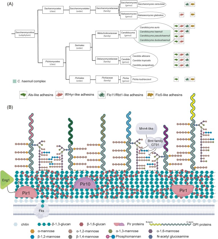
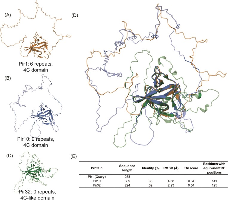
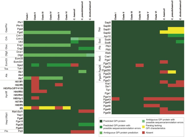
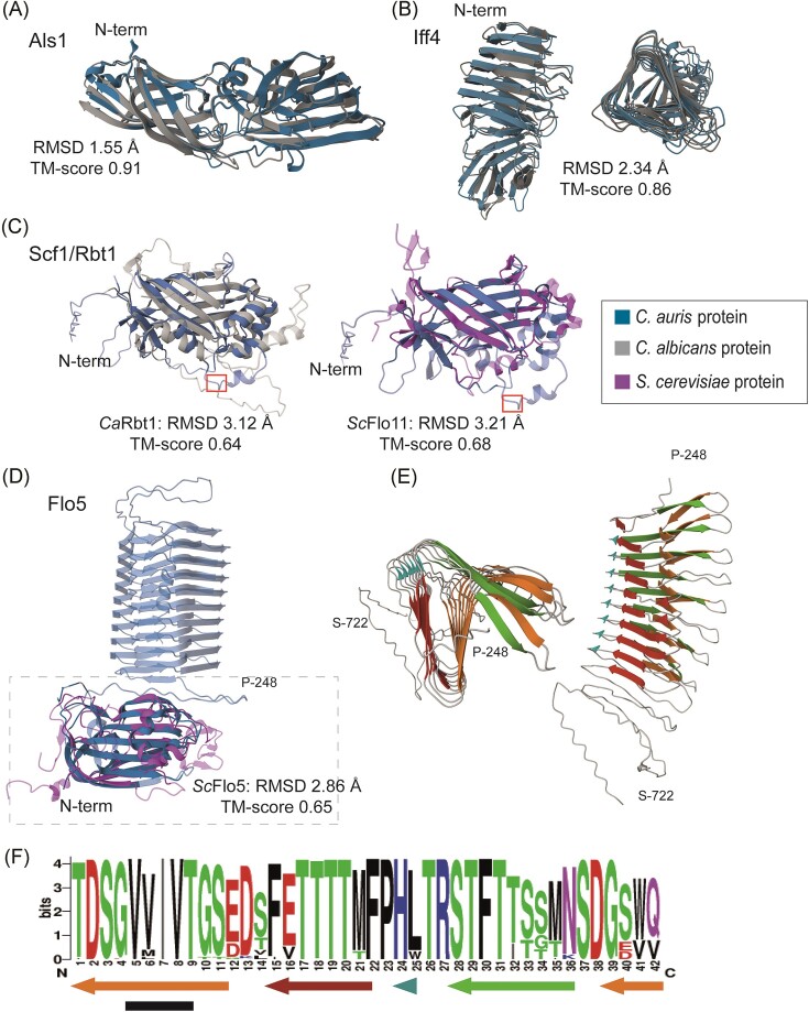
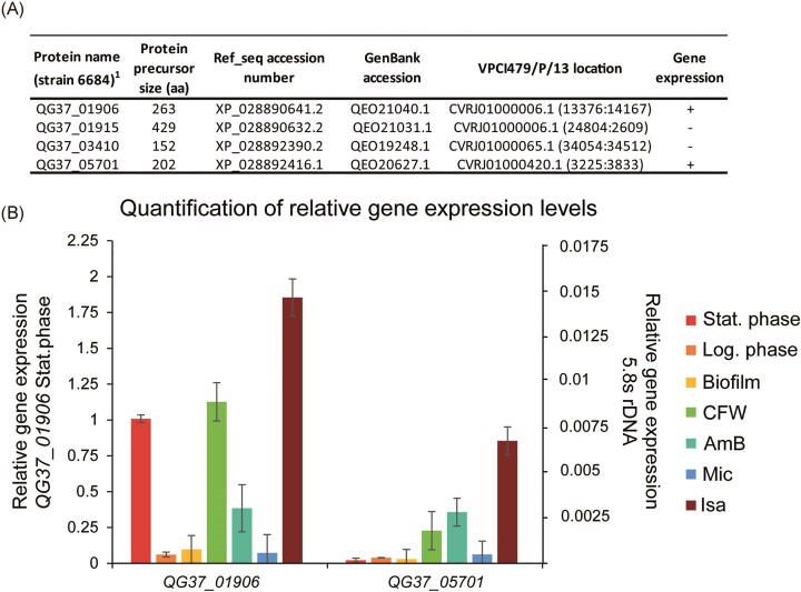
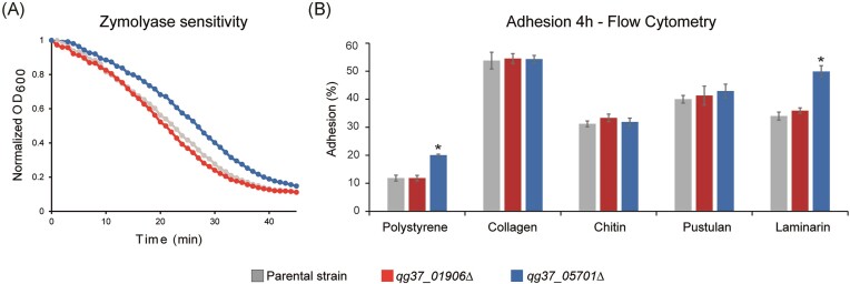
The drug-resistant pathogenic yeast Candidozyma auris (formerly named Candida auris) is considered a critical health problem of global importance. As the cell wall plays a crucial role in pathobiology, here we performed a detailed bioinformatic analysis of its biosynthesis in C. auris and related Candidozyma haemuli complex species using Candida albicans and Saccharomyces cerevisiae as references. Our data indicate that the cell wall architecture described for these reference yeasts is largely conserved in Candidozyma spp.; however, expansions or reductions in gene families point to subtle alterations, particularly with respect to β--1,3--glucan synthesis and remodeling, phosphomannosylation, β-mannosylation, and glycosylphosphatidylinositol (GPI) proteins. In several aspects, C. auris holds a position in between C. albicans and S. cerevisiae, consistent with being classified in a separate genus. Strikingly, among the identified putative GPI proteins in C. auris are adhesins typical for both Candida (Als and Hyr/Iff) and Saccharomyces (Flo11 and Flo5-like flocculins). Further, 26 putative C. auris GPI proteins lack homologs in Candida genus species. Phenotypic analysis of one such gene, QG37_05701, showed mild phenotypes implicating a role associated with cell wall β-1,3-glucan. Altogether, our study uncovered a wealth of information relevant for the pathogenicity of C. auris as well as targets for follow-up studies.

Keywords: Candida auris; GPI proteins; adhesins; candidiasis; cell wall; glucan; mannosylation.


Conflict of interest statement

None declared.

Figures

Figure 1.
Figure 1.
Genomic analysis of cell wall biosynthesis in C. auris. (A) Taxonomic tree including the species analyzed in this study and the most important causal agents of candidiasis according to the WHO (World Health Organization 2022). Putative adhesin families present in each of the species are indicated on the right. (B) Proposed cell wall architectural model based on genomic data. Major distinctive features explained in the text are indicated. Colors of N-terminal parts of GPI proteins represent different protein families (including adhesins).
Figure 2.
Figure 2.
3D structural analysis of C. auris Pir proteins. (A–B) Pir1 paralogs harboring internal repeats and the conserved 4C domain. (C) Pir32 paralog lacking repeats and harboring a modified 4C domain. (D) RCSB TM-alignment of the three proteins showing structural conservation of the 4C domain. (E) Details of sequence and tertiary structure similarity between Pir1 and the other two Pir family proteins.
Figure 3.
Figure 3.
Conserved putative GPI proteins in the six C. auris clades and in C. haemuli complex species. GPI proteins were identified in silico and annotated according to their closest homologs in C. albicans or S. cerevisiae Flo5, as detailed in the “Materials and Methods” section.
Figure 4.
Figure 4.
Comparative structural analysis of C. auris adhesins. (A–D) Cartoon presentations of modeled three-dimensional structures (AlphaFold2) of putative ligand-binding domains of C. auris adhesins aligned (RCSB TM-alignment) with their closest C. albicans or S. cerevisiae homologs. (A) Als family protein Als1; (B) Iff/Hyr family protein Iff4, side and top views; and (C) Scf1/Rbt1. A canonical surface-exposed Kex2 cleavage site (KR/DV) at positions 216–219 in Scf1 is indicated by a red box. (D) Flo5 (strain B11220). The Flo5 model includes the repeat domain (top part) downstream of the ligand-binding domain (boxed), the latter aligned to ScFlo5. N-terminal residues in the structures of the C. auris proteins are indicated. (E) Top and side view of the strain B11220 Flo5 42-aa repeat domain (aa 248–722). (F) Sequence logo of the 42-aa repeat based on all Flo5 repeats in six representative strains (one per clade). Arrows indicate β-sheet forming regions, and the black bar marks a small region with high β-aggregation propensities according to TANGO. Ca, C. albicans and Sc, S. cerevisiae.
Figure 5.
Figure 5.
qPCR gene expression analysis of C. auris GPI protein-encoding genes lacking homologs in the Candida genus. (A) Four GPI protein-encoding genes were selected for gene expression analysis. (B) Relative gene expression levels of the two actively transcribed genes QG37_01 906 and QG37_05701. Data were normalized to 5.8S rDNA expression and plotted in comparison to QG37_01 906 (left axis) and 5.8S rDNA (right axis) transcript levels at the stationary phase. Shown are average values and standard deviations of two biological samples measured in triplicate.
Figure 6.
Figure 6.
Deletion of QG37_05 701 but not QG37_01 906 affects glucan-related and adhesion properties of C. auris. (A) Zymolyase sensitivity. (B) Adhesion assays. Percentage of cells adhering to polystyrene (PS) and PS coated with different biomolecules after 4 h of incubation, measured by flow cytometry. Details concerning the number of replicates in each assay and statistical analysis are provided in the “Materials and Methods” section. Error bars indicate standard deviations. P-values < .05 (*) were considered statistically significant.

Similar articles

Cited by

References

    1. Alvarado M, Bartolome Alvarez J, Lockhart SR et al. Identification of Candida auris and related species by multiplex PCR based on unique GPI protein-encoding genes. Mycoses. 2021;64:194–202. - PMC - PubMed
    1. Alvarado M, Gomez-Navajas JA, Blazquez-Munoz MT et al. Integrated post-genomic cell wall analysis reveals floating biofilm formation associated with high expression of flocculins in the pathogen Pichia kudriavzevii. PLoS Pathog. 2023;19:e1011158. - PMC - PubMed
    1. Arendrup MC, Prakash A, Meletiadis J et al. Comparison of EUCAST and CLSI Reference microdilution MICs of eight antifungal compounds for Candida auris and associated tentative epidemiological cutoff values. Antimicrob Agents Chemother. 2017;61:e00485–17. - PMC - PubMed
    1. Arensman K, Miller JL, Chiang A et al. Clinical outcomes of patients treated for Candida auris infections in a multisite health system, Illinois, USA. Emerg Infect Dis. 2020;26:876–80. - PMC - PubMed
    1. Bader O, Krauke Y, Hube B. Processing of predicted substrates of fungal Kex2 proteinases from Candida albicans, C. glabrata, Saccharomyces cerevisiae and Pichia pastoris. BMC Microbiol. 2008;8:116. - PMC - PubMed

Publication types

MeSH terms