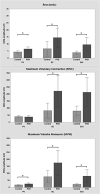
Pelvic floor muscle activation amplitude at rest, during voluntary contraction, and during Valsalva maneuver-a comparison between those with and without provoked vestibulodynia
- PMID: 39657059
- PMCID: PMC12001039
- DOI: 10.1093/jsxmed/qdae170
Pelvic floor muscle activation amplitude at rest, during voluntary contraction, and during Valsalva maneuver-a comparison between those with and without provoked vestibulodynia
Abstract
Background: The neuromuscular contribution to increased tone of the pelvic floor muscles (PFMs) observed among those with provoked vestibulodynia (PVD) is unclear.
Aim: To determine if PFM activity differs between those with provoked PVD and pain free controls, and if the extent of PFM activation at rest or during activities is associated with pain sensitivity at the vulvar vestibule, psychological, and/or psychosexual outcomes.
Methods: This observational case-control study included forty-two volunteers with PVD and 43 controls with no history of vulvar pain. Participants completed a series of questionnaires to evaluate pain, pain catastrophizing, depression, anxiety and stress, and sexual function, then underwent a single laboratory-based assessment to determine their pressure pain threshold at the vulvar vestibule and electromyographic (EMG) signal amplitudes recorded from three PFMs (pubovisceralis, bulbocavernosus, and external anal sphincter).
Outcomes: EMG signal amplitude recorded at rest, during maximum voluntary contraction (MVC), and during maximal effort Valsalva maneuver, pressure pain threshold at the vulvar vestibule, and patient-reported psychological (stress, anxiety, pain catastrophizing, central sensitization) and psychosexual (sexual function) outcomes.
Results: Participants with PVD had higher activation compared to controls in all PFMs studied when at rest and during Valsalva maneuver. There were no group differences in EMG amplitude recorded from the pubovisceralis during MVC (Cohen's d = 0.11), but greater activation was recorded from the bulbocavernosus (d = 0.67) and the external anal sphincter(d = 0.54) among those with PVD. When EMG amplitudes at rest and on Valsalva were normalized to activation during MVC, group differences were no longer evident, except at the pubovisceralis, where tonic EMG amplitude was higher among those with PVD (d = 0.42). While those with PVD had lower vulvar pressure pain thresholds than controls, there were no associations between PFM EMG amplitude and vulvar pain sensitivity nor psychological or psychosexual problems.
Clinical implications: Women with PVD demonstrate evidence of PFM overactivity, yet the extent of EMG activation is not associated with vulvar pressure pain sensitivity nor psychological/psychosexual outcomes. Interventions aimed at reducing excitatory neural drive to these muscles may be important for successful intervention.
Strengths and limitations: This study includes a robust analysis of PFM EMG. The analysis of multiple outcomes may have increased the risk statistical error, however the results of hypothesis testing were consistent across the three PFMs studied. The findings are generalizable to those with PVD without vaginismus.
Conclusions: Those with PVD demonstrate higher PFM activity in the bulbocavernosus, pubovisceralis, and external anal sphincter muscles at rest, during voluntary contraction (bulbocavernosus and external anal sphincter) and during Valsalva maneuver; yet greater activation amplitude during these tasks is not associated with greater vulvar pressure pain sensitivity nor psychological or psychosexual function.
Keywords: electromyography; muscle tone; overactivity; pelvic floor; pelvic pain; provoked vestibulodynia; vulvodynia.
© The Author(s) 2024. Published by Oxford University Press on behalf of The International Society for Sexual Medicine.
Conflict of interest statement
Linda McLean is a Scientific Advisor for Cntrl+ Inc. (Canda) and Freyya Inc. (United States).
Figures


References
-
- Harlow BL, Steward EG. A population-based assessment of chronic unexplained vulvar pain: have we underestimated the prevalence of vulvodynia? J American Medical Women’s Association. 2003;58:82–88. - PubMed
Publication types
MeSH terms
Grants and funding
LinkOut - more resources
Full Text Sources

