. 2025 Jan 21;219(1):uxae117.
doi: 10.1093/cei/uxae117.

FACS-based detection of extracellular ASC specks from NLRP3 inflammasomes in inflammatory diseases

Affiliations

FACS-based detection of extracellular ASC specks from NLRP3 inflammasomes in inflammatory diseases

Joanne Topping et al. Clin Exp Immunol. .

Abstract

Introduction: The apoptosis-associated speck-like protein containing a caspase recruitment domain (ASC) is crucial for inflammasome assembly and activation of several inflammasomes, including NLRP3. ASC aggregates are detected in human sera post pyroptotic cell death, but their inflammasome origin remains unclear.

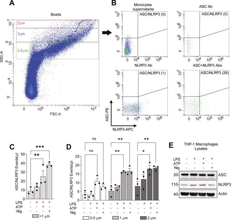
Method: This study aimed to develop a method to detect ASC aggregates originating from NLRP3 inflammasomes. Initially, human monocytes, macrophages, and THP-1 ASC reporter cells were employed to validate the detection of ASC/NLRP3-positive events through flow cytometry.

Results: The presence of ASC/NLRP3 specks was confirmed in cell supernatants from monocytes and macrophages treated with LPS and nigericin or ATP. Flow cytometry analysis identified double-positive specks in patient sera from inflammatory conditions when compared with healthy controls. Elevated ASC/NLRP3 specks were observed in conditions such as cryopyrin-associated periodic syndrome and Schnitzler's syndrome.

Conclusion: We validated fluorescence-activated cell sorting as a reliable method for detecting ASC/NLRP3 specks in human sera, with potential diagnostic and monitoring applications in certain systemic autoinflammatory diseases.

Keywords: ASC; NLRP3; SAID; inflammasome; inflammation.


Conflict of interest statement

S.S. received honoraria for participation in advisory board meetings and speaking engagements, travel support, and research grants from SOBI and Novartis. Other authors have no conflicts of interest to declare.

Figures

Graphical Abstract
Graphical Abstract
Figure 1:
Figure 1:
Extracellular ASC in THP-1 cells. (A) Detection of ASC specks in THP-1-defASC or THP-1-ASC-GFP by fluorescent microscopy, stimulated with LPS, LPS + ATP, or LPS + nigericin (LPS + Nig) as indicated in the methods. Each dotted square is shown on the right of the panel as magnified. n = 3 independent experiments were carried out. (B) Extracellular ASC was imaged in the CD7; the images presented here are representative of n = 3 independent experiments with 10 images each. (C) Immunoblots of lysates or supernatants of THP-1-defASC or THP-1-ASC-GFP cells treated as indicated. Images are representative of n = 3 independent experiments. (D and E) ASC specks from THP-1-defASC or THP-1-ASC-GFP cells were captured using flow cytometry, with gating around the 1-μm bead size, showing the ASC events/μl in 30 μl of total volume. (F) ELISA assays were used to detect IL-1β in the supernatants of the cells. P-values *≤0.05, **≤0.01, ***≤0.001 from two‐way ANOVA following adjustment for multiple comparisons.
Figure 2:
Figure 2:
ASC specks validation on THP-1 cells. (A–C) Detection of ASC specks in THP-1-defASC or THP-1-ASC-GFP by FACS, stimulated with LPS, LPS + Nig, or using the indicated control, as described in the methods. Validation of ASC specks using flow cytometry with a gating around the 0.5-,1-, and 2-μm bead size, showing the ASC events/μl in 30 μl of total volume. The number next to the ASC specks represents the total number of events. A total of six independent experiments were carried out.
Figure 3:
Figure 3:
Secretion of ASC/NLRP3 by macrophages. (A and B) Detection of ASC/NLRP3 specks in the supernatant of primary human monocytes, stimulated as shown in the figure, using flow cytometry with a gating around the 0.5-,1-, and 2-μm bead size, showing the ASC/NLRP3 events/μl in 30 μl of total volume. (C) Immunoblots of lysates of primary human monocytes then treated as indicated, immunoblots are representative of n = 3 independent experiments. (D) Detection of ASC/NLRP3 specks in the supernatant of MDMs, stimulated as shown in the figure and transfected with siRNA against NLRP3 or non-targeting control (CTRL). (E) Immunoblots of lysates of MDMs treated as indicated. Immunoblots are representative of n = 3 independent experiments. P-values *≤0.05, **≤0.01, ***≤0.001 from two‐way ANOVA following adjustment for multiple comparisons.
Figure 4:
Figure 4:
ASC/NLRP3 specks on monocytes and THP-1 macrophages. (A and B) Validation of the detection of ASC/NLRP3 specks in human monocytes by FACS; the indicated control is as shown in the figure. Three independent experiments were carried out. Validation of ASC specks using flow cytometry with gating around the 0.5-,1-, and 2-μm bead size, showing the ASC/NLRP3 events/μl in 30 μl of total volume. The number next to ASC/NLRP3 specks represents the total number of events. (C) ASC/NLRP3 specks in human monocytes representing the events/μl in 30 μl of total volume combining the events of 1- and 2-μm gates. (D) Detection of ASC/NLRP3 specks in the supernatant of THP-1 macrophages, stimulated as shown in the figure, using flow cytometry with a gating around the 0.5-,1-, and 2-μm bead size, showing the ASC/NLRP3 events/μl in 30 μl of total volume. (E) Immunoblots of lysates of THP-1 macrophages then treated as indicated. Immunoblots are representative of n = 3 independent experiments. P-values *≤0.05, **≤0.01, ***≤0.001 from two‐way ANOVA following adjustment for multiple comparisons.
Figure 5:
Figure 5:
Detection on ASC/NLRP3 in sera of patients with inflammatory disorders. (A and B) Detection of ASC/NLRP3 specks in the sera of HCs (n = 6), SAIDs (n = 7), and CF (n = 3) patients. Events were recorded using flow cytometry with a gating around the 1- and 2-μm bead size, showing the ASC/NLRP3 events/μl in 30 μl of total volume. (C) Detection of ASC/NLRP3 specks in sera from HC (n = 32), CAPS (n = 7), Schnitzler’s syndrome (n = 7), and FMF (n = 15) using FACS-based methodology described above. (D) Independent replicates of 11 serum samples analysed on two dates, more than 4 weeks apart. Samples were aliquoted and frozen at −80°C before analysis. Dots show the mean of duplicate repeats from one frozen aliquot; lines connect the means of the same sample. To look for significant difference between the groups. The Kruskal–Wallis test with Dunn’s multiple comparison test was performed, or a Wilcoxon matched-pairs signed rank test was used P-values *≤0.05, **≤0.01, ***≤0.001.

Similar articles

References

    1. Broz P, Dixit VM.. Inflammasomes: mechanism of assembly, regulation and signalling. Nat Rev Immunol 2016, 16, 407–20. doi: https://doi.org/10.1038/nri.2016.58 - DOI - PubMed
    1. Cheng J, Waite AL, Tkaczyk ER, Ke K, Richards N, Hunt AJ, et al. Kinetic properties of ASC protein aggregation in epithelial cells. J Cell Physiol 2010, 222, 738–47. doi: https://doi.org/10.1002/jcp.22005 - DOI - PMC - PubMed
    1. Hoss F, Rodriguez-Alcazar JF, Latz E.. Assembly and regulation of ASC specks. Cell Mol Life Sci 2017, 74, 1211–29. doi: https://doi.org/10.1007/s00018-016-2396-6 - DOI - PMC - PubMed
    1. Lara-Reyna S, Caseley EA, Topping J, Rodrigues F, Jimenez Macias J, Lawler SE, et al. Inflammasome activation: from molecular mechanisms to autoinflammation. Clin Transl Immunol 2022, 11, e1404. doi: https://doi.org/10.1002/cti2.1404 - DOI - PMC - PubMed
    1. Franklin BS, Bossaller L, De Nardo D, Ratter JM, Stutz A, Engels G, et al. The adaptor ASC has extracellular and ‘prionoid’ activities that propagate inflammation. Nat Immunol 2014, 15, 727–37. doi: https://doi.org/10.1038/ni.2913 - DOI - PMC - PubMed

MeSH terms

Substances