. 2025 Jan 11;53(2):gkae1207.
doi: 10.1093/nar/gkae1207.

Novel BRCA1-PLK1-CIP2A axis orchestrates homologous recombination-mediated DNA repair to maintain chromosome integrity during oocyte meiosis

Affiliations

Novel BRCA1-PLK1-CIP2A axis orchestrates homologous recombination-mediated DNA repair to maintain chromosome integrity during oocyte meiosis

Crystal Lee et al. Nucleic Acids Res. .

Abstract

Double-strand breaks (DSBs) are a formidable threat to genome integrity, potentially leading to cancer and various genetic diseases. The prolonged lifespan of mammalian oocytes increases their susceptibility to DNA damage over time. While somatic cells suppress DSB repair during mitosis, oocytes exhibit a remarkable capacity to repair DSBs during meiotic maturation. However, the precise mechanisms underlying DSB repair in oocytes remain poorly understood. Here, we describe the pivotal role of the BRCA1-PLK1-CIP2A axis in safeguarding genomic integrity during meiotic maturation in oocytes. We found that inhibition of homologous recombination (HR) severely impaired chromosome integrity by generating chromosome fragments during meiotic maturation. Notably, HR inhibition impaired the recruitment of CIP2A to damaged chromosomes, and the depletion of CIP2A led to chromosome fragmentation following DSB induction. Moreover, BRCA1 depletion impaired chromosomal recruitment of CIP2A, but not vice versa. Importantly, the impaired chromosomal recruitment of CIP2A could be rescued by PLK1 inhibition. Consequently, our findings not only underscore the importance of the chromosomal recruitment of CIP2A in preventing chromosome fragmentation, but also demonstrate the regulatory role of the BRCA1-PLK1-CIP2A axis in this process during oocyte meiotic maturation.


Figures

Graphical Abstract
Graphical Abstract
Figure 1.
Figure 1.
RAD51 inhibition using small molecule B02 impairs HR and DSB repair in oocytes. Oocytes at the MI stage were treated with ETP for 15 min with or without B02. Control oocytes were treated with DMSO. (A) Representative images of MI chromosomes stained with RAD51 and ACA antibodies. Scale bar, 10 μm. In the bottom panel of the DMSO control (enlarged), centromeres are highlighted with a dashed box and further magnified in the right panel. (B) Quantification of RAD51 intensity. (C) MI oocytes were treated with ETP for 15 min, washed out and cultured for 1 h to allow recovery from DNA damage. (D) Quantification of TUNEL intensity. Data are presented as the mean ± standard error of the mean (SEM) from three independent experiments. ****P < 0.0001, ***P < 0.0006; ns, not significant.
Figure 2.
Figure 2.
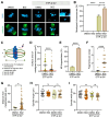
Inhibition of HR during meiotic maturation causes chromosome fragmentation following DSB induction. GV oocytes were treated with ETP for 15 min, washed out and matured to the MII stage in the presence of B02 for up to 15 h. Control oocytes were treated with DMSO instead of ETP and matured with or without B02. (A) Representative images of MII oocytes showing chromosome and spindle organization. Chromosome fragments in B02-treated oocytes are boxed and enlarged. Scale bar, 10 μm. (B) Quantification of the number of oocytes with misaligned chromosomes. (C) Schematic diagram of the parameters measured in the graphs (D–I). (DG) Quantification of the surface area of misaligned chromosomes, MII fragmentation, number of fragments per oocyte and the distance of fragments from the center of the metaphase plate. (H, I) Quantification of spindle length and width. Data are presented as mean ± SEM of three independent experiments. ****P < 0.0001, *P < 0.03; ns, not significant.
Figure 3.
Figure 3.
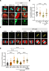
HR inhibition impairs the chromosomal association of CIP2A and disrupts chromosome conformation. GV oocytes were treated with ETP for 15 min, washed out and matured in the presence of B02 for up to 15 h. (A) Representative images of MII chromosomes stained with CIP2A and ACA antibodies. Scale bar, 10 μm. (B) Quantification of overall CIP2A intensity. (C) Enlarged views of the boxed regions (a–e) in panel (A). Chromosomes are categorized into three groups: intact, fragmented and aggregated. Centric and acentric fragments are marked with arrows and arrowheads, respectively. (D, E) Quantification of CIP2A-positive chromatid and CIP2A intensity on each fragment in ETP-treated groups. (F) Quantification of the types of chromosome conformations observed in each treatment group. (G) Distribution of centric and acentric fragments in B02-treated oocytes with DNA damage. Data are presented as mean ± SEM of three independent experiments. ****P< 0.0001.
Figure 4.
Figure 4.
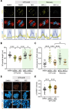
CIP2A recruitment onto damaged chromosomes is dependent on the HR pathway. Oocytes at the MI stage were treated with ETP in the presence of either DMSO or B02 for 15 min. After ETP washout, oocytes were allowed to recover for 1 h in the presence (+) or absence (−) of B02. (A) Representative images of MI oocytes showing CIP2A chromosomal recruitment. Profiling graphs are shown below to illustrate the pole-to-chromosome distribution of CIP2A. Scale bar, 10 μm. (B, C) Quantification of MI spindle length and chromosomal CIP2A intensity. (D) Representative images of MI chromosomes stained with p-TOPBP1 and ACA antibodies. Scale bar, 10 μm. (E) Quantification of p-TOPBP1 intensity. Data are presented as mean ± SEM of three independent experiments. ****P< 0.0001; ns, not significant.
Figure 5.
Figure 5.
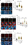
Chromosomal recruitment of CIP2A is independent of NHEJ, MMEJ and RAD52-dependent repair pathways. (A) Representative images of MI oocytes treated with ETP for 15 min in the presence of SCR7 (NHEJ inhibitor) or ART558 (MMEJ inhibitor). Oocytes were subjected to immunostaining with CIP2A antibody, showing chromosomal recruitment of CIP2A. Scale bar, 10 μm. (B, C) Quantification of MI spindle length and chromosomal CIP2A intensity. Data are presented as mean ± SEM of three independent experiments. ****P< 0.0001; ns, not significant. (D) Representative images of MI oocytes treated with ETP for 15 min in the presence of D-I03 (RAD52 inhibitor). Oocytes were subjected to immunostaining with CIP2A antibody, showing chromosomal recruitment of CIP2A. Scale bar, 10 μm. (E) Quantification of chromosomal CIP2A intensity. Data are presented as mean ± SEM of three independent experiments. ****P< 0.0001; ns, not significant.
Figure 6.
Figure 6.
CIP2A depletion leads to chromosome fragmentation after DSB induction. (A) Scheme of Trim-away experiments to acutely deplete CIP2A in oocytes at the MI stage. GV oocytes were microinjected with Trim21-mCherry mRNA and cultured for 8 h to reach the MI stage. After confirming mCherry expression, MI oocytes were injected with either IgG control antibody (CTL) or CIP2A antibody (CIP2A-TA). After 1 h incubation to allow CIP2A depletion, oocytes were treated with ETP for 15 min, washed out and matured to the MII stage. (B) Representative images of MII oocytes showing chromosome fragmentations and CIP2A intensity. Scale bar, 10 μm. (C, D) Quantification of chromosomal CIP2A intensity and chromosome fragmentation. Data are presented as mean ± SEM of three independent experiments. ****P< 0.0001. (E) Correlation of CIP2A intensity against chromosome fragmentation after ETP treatment. r = −0.9091, P< 0.05. (F) Representative images of MII chromosomes illustrating chromosome fragmentation and aggregation. Centric and acentric fragments are marked with arrows and arrowheads, respectively. Scale bar, 10 μm. (G) Quantification of the types of chromosome conformations observed in each treatment group. (H) Distribution of centric and acentric fragments in CIP2A-depleted oocytes with DNA damage. Data are presented as mean ± SEM of three independent experiments.
Figure 7.
Figure 7.
BRCA1 depletion impairs chromosomal recruitment of CIP2A induced by DNA damage. (A) Oocytes at the MI stage were treated with ETP in the presence of either DMSO or B02 for 15 min. Representative images of MI chromosomes stained with BRCA1 antibody. Scale bar, 10 μm. (B) Quantification of BRCA1 intensity. (C) Representative images of CIP2A chromosomal recruitment in MI oocytes after BRCA1 Trim-away (BRCA1-TA) and ETP treatment. BRCA1 Trim-away was conducted as described for CIP2A Trim-away. Scale bar, 10 μm. (D) Quantification of chromosomal CIP2A intensity. (E) Representative images of MI chromosomes stained with BRCA1 antibody after CIP2A Trim-away (CIP2A-TA). Scale bar, 10 μm. (F) Quantification of BRCA1 intensity. Data are presented as mean ± SEM of three independent experiments; ****P< 0.0001; ns, not significant.
Figure 8.
Figure 8.
Impaired chromosomal recruitment of CIP2A induced by HR inhibition is restored by PLK1 inhibition. (A) Representative images of p-T210-PLK1 at spindle poles of MI oocytes after BRCA1 Trim-away (BRCA-TA). Scale bar, 10 μm. (B) Quantification of p-T210-PLK1 intensity at spindle poles. (C) Representative images of oocytes showing CIP2A chromosomal recruitment after treatment with ETP, B02 and BI2536. Scale bar, 10 μm. (D) Quantification of chromosomal CIP2A intensity. Data are presented as mean ± SEM of three independent experiments. ****P< 0.0001, ***P< 0.0006.

Similar articles

Cited by

References

    1. Jackson S.P., Bartek J.. The DNA-damage response in human biology and disease. Nature. 2009; 461:1071–1078. - PMC - PubMed
    1. Shrivastav M., De Haro L., Nickoloff J.A.. Regulation of DNA double-strand break repair pathway choice. Cell Res. 2008; 18:134–147. - PubMed
    1. Ceccaldi R., Rondinelli B., D’Andrea A.D.. Repair pathway choices and consequences at the double-strand break. Trends Cell Biol. 2016; 26:52–64. - PMC - PubMed
    1. Rogakou E.P., Pilch D.R., Orr A.H., Ivanova V.S., Bonner W.M.. DNA double-stranded breaks induce histone H2AX phosphorylation on serine 139. J. Biol. Chem. 1998; 273:5858–5868. - PubMed
    1. Leem J., Lee C., Choi D.Y., Oh J.S.. Distinct characteristics of the DNA damage response in mammalian oocytes. Exp. Mol. Med. 2024; 56:319–328. - PMC - PubMed

MeSH terms

LinkOut - more resources