. 2025 Feb;206(2):666-674.
doi: 10.1111/bjh.19945. Epub 2024 Dec 10.

Imaging flow cytometry as a novel approach for the diagnosis of heparin-induced thrombocytopenia

Affiliations

Imaging flow cytometry as a novel approach for the diagnosis of heparin-induced thrombocytopenia

Julie Carré et al. Br J Haematol. 2025 Feb.

Abstract

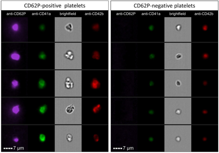
Heparin-induced thrombocytopenia (HIT) is an adverse reaction characterized by anti-PF4-heparin antibody generation and hypercoagulability. Imaging flow cytometry (IFC) provides a detailed morphological analysis of platelets, which change upon activation. We evaluated IFC-derived morphometric features to detect platelet activation and developed a functional assay for HIT diagnosis. We analysed blood samples from 42 patients with suspected HIT and extracted platelet size, shape and texture features using IFC. The morphological features were compared with CD62P expression, light transmission aggregometry (LTA) and a serotonin release assay (SRA) in terms of their ability to predict a HIT diagnosis. Five IFC-derived morphological features (area, circularity, contrast, diameter and major axis) significantly distinguished resting from activated platelets. The major axis feature performed best for HIT diagnosis, with a sensitivity of 89.3% and a specificity of 92.9% versus functional assays (LTA/SRA); this diagnostic performance was similar to that of CD62P expression on the same platelet donors. The area and diameter had similar specificity (92.9%) and a slightly lower sensitivity (85.7%). The morphological features associated with platelet activation might be effective markers for the diagnosis of HIT, matching platelet CD62P expression assay performance. The high-throughput IFC exploration of platelet activation offers new perspectives in label-free analysis and time-saving.

Keywords: heparin‐induced thrombocytopenia; imaging flow cytometry; laboratory diagnosis.


Conflict of interest statement

The authors declare no conflicts of interest.

Figures

FIGURE 1
FIGURE 1
Illustrations of TRAP‐6‐activated, CD62P‐positive platelets (left panel) and resting, CD62P‐negative platelets (right panel). The platelets were viewed in the bright‐field channel and in the CD62‐BV421, CD41a‐FITC and CD42b APC channels.
FIGURE 2
FIGURE 2
The ability of morphometric features to distinguish between resting and activated platelets. Using IFC, the mean variations in each morphometric feature (area (A), circularity (B), contrast (C), diameter (D), major axis (E) and modulation (F)) were measured in the baseline resting state (control) and the TRAP‐6‐activated state (n = 21). ****: P‐value <0.0001, ***: P‐value <0.001, ns, not significant.
FIGURE 3
FIGURE 3
The value of IFC‐derived morphometric features for HIT diagnosis. Variations in morphometric features (area: A–B, circularity: C–D, contrast: E–F, diameter: G–H and major axis: I–J, modulation: K–L) with low‐dose UFH are shown on the left (A, C, E, G, I and K), and those with high‐dose UFH are shown on the right (B, D, F, H, J and L) (n = 28 positive HIT patients tested once and n = 14 negative HIT patients tested twice). True positives and negatives are shown as filled circles, whereas false positives and negatives are shown as open squares or triangles respectively. The dashed lines indicate the positivity threshold. ****: P‐value <0.0001, ns: not significant.
FIGURE 4
FIGURE 4
Comparison of the CD62P positivity rates at various heparin concentrations for patients diagnosed with HIT versus patients without HIT. The bee‐swarm plot illustrates the percentage of platelets expressing CD62P for (A) patients diagnosed with HIT versus (B) patients without HIT at various UFH concentrations (H0, H1, H500) (n = 28 positive HIT patients tested once and n = 14 negative HIT patients tested twice). True positives and negatives are represented by filled circles, whereas false positives and negatives are shown as open squares or triangles respectively. The dashed lines represent the positivity threshold. H0: No UFH; H1: A UFH concentration of 1 IU/mL; H500: A UFH concentration of 500 IU/mL. ****: P‐value <0.0001, ***: P‐value <0.001, **: P‐value <0.01, *: P‐value <0.05, ns, not significant.
FIGURE 5
FIGURE 5
Reproducibility of HIT diagnosis using morphometric features across diverse platelet donors. Variations in morphometric features (area: A, B, circularity: C, D, contrast: E, F, diameter: G, H, major axis: I, J and modulation: K, J) with low‐dose UFH are shown on the left (A, C, E, G, I and K), and those with high‐dose UFH are shown on the right (B, D, F, H, J and L) (n = 6). All six platelet donors were tested using the same plasma from HIT patient number 1. Each random platelet donor is shown as a colour‐filled symbol. True positives are shown as filled circles, whereas false negatives are shown as filled triangles. The star symbol corresponds to the good responder platelet donor used within Figure 3. The dashed lines indicate the positivity threshold.

References

    1. Arepally GM. Heparin‐induced thrombocytopenia. Blood. 2017;129(21):2864–2872. - PMC - PubMed
    1. Arepally GM, Padmanabhan A. Heparin‐induced thrombocytopenia: a focus on thrombosis. Arterioscler Thromb Vasc Biol. 2021;41(1):141–152. - PMC - PubMed
    1. Cuker A, Arepally G, Crowther MA, Rice L, Datko F, Hook K, et al. The HIT expert probability (HEP) score: a novel pre‐test probability model for heparin‐induced thrombocytopenia based on broad expert opinion. J Thromb Haemost. 2010;8(12):2642–2650. - PubMed
    1. Nagler M, Bachmann LM, Ten Cate H, Ten Cate‐Hoek A. Diagnostic value of immunoassays for heparin‐induced thrombocytopenia: a systematic review and meta‐analysis. Blood. 2016;127(5):546–557. - PubMed
    1. Sun L, Gimotty PA, Lakshmanan S, Cuker A. Diagnostic accuracy of rapid immunoassays for heparin‐induced thrombocytopenia: a systematic review and meta‐analysis. Thromb Haemost. 2016;115(5):1044–1055. - PubMed