Differentiating mycosis fungoides lesions from their mimickers clinically and histologically: A single tertiary center retrospective analysis in Saudi Arabia
- PMID: 39658107
- PMCID: PMC11629653
- DOI: 10.15537/smj.2024.45.12.20240796
Differentiating mycosis fungoides lesions from their mimickers clinically and histologically: A single tertiary center retrospective analysis in Saudi Arabia
Abstract
Objectives: To identify the clinical and histological features of MF that can assist in distinguishing MF from MF-mimicking cases. Although mycosis fungoides (MF) is the most common subtype of cutaneous T-cell lymphoma, clinicopathological correlations are required to establish an accurate diagnosis, which are currently lacking.
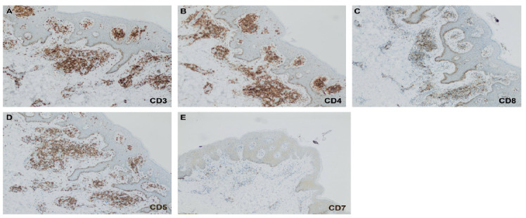
Methods: This retrospective observational study evaluated the clinical presentations, characteristics, and histological features of 56 patients with suspected MF who presented to our clinic between January 2018 and August 2022. Immunohistochemistry was performed, and the loss of CD5 and CD7 T-cells and T-cell receptor rearrangement was evaluated.
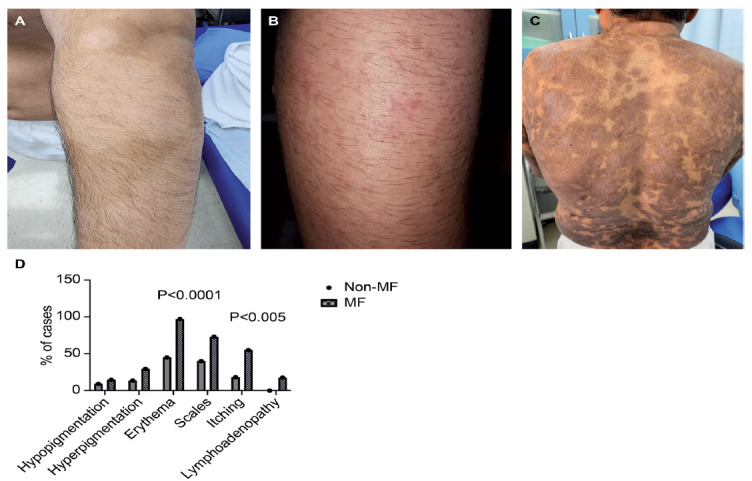
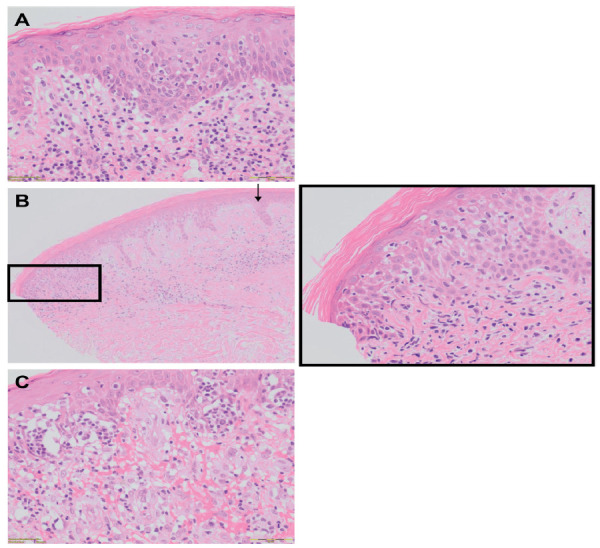
Results: Overall, 34 patients were diagnosed with MF, whereas 22 were not. Clinical erythroderma, poikiloderma, and nodular presentations were more commonly associated with a histological diagnosis of MF than macular presentations. Erythema and pruritus were significantly more common in MF cases than in MF-mimicking cases (p<0.05). Epidermotropism and parakeratosis were the key histological features for diagnosing MF. Additionally, Pautrier's microabscesses correlated with the clinical presentation of plaques in MF. Loss of CD7 expression on the T-cell surface was observed even in early-stage MF cases.
Conclusion: Our proposed diagnostic features are statistically valid and, along with those previously reported, can aid in identifying and distinguishing MF cases from MF-mimicking cases.
Keywords: dermatology; histopathology; immunohistochemistry; mycosis fungoides.
Copyright: © Saudi Medical Journal.
Figures
References
-
- Hossain C, Jennings T, Duffy R, Knoblauch K, Gochoco A, Chervoneva I, et al. The histological prevalence and clinical implications of folliculotropism and syringotropism in mycosis fungoides. Chin Clin Oncol 2019; 8: 6. - PubMed
-
- Willemze R, Jaffe ES, Burg G, Cerroni L, Berti E, Swerdlow SH, et al. WHO-EORTC classification for cutaneous lymphomas. Blood 2005; 105: 3768–3785. - PubMed
-
- Wilcox RA. Cutaneous T-cell lymphoma: 2017 update on diagnosis, risk-stratification, and management. Am J Hematol 2017; 92: 1085–1102. - PubMed
-
- Quaglino P, Pimpinelli N, Berti E, Calzavara-Pinton P, Alfonso Lombardo G, Rupoli S, et al. Time course, clinical pathways, and long-term hazards risk trends of disease progression in patients with classic mycosis fungoides: a multicenter, retrospective follow-up study from the Italian Group of Cutaneous Lymphomas. Cancer 2012; 118: 5830–5839. - PubMed
-
- Pimpinelli N, Olsen EA, Santucci M, Vonderheid E, Haeffner AC, Stevens S, et al. Defining early mycosis fungoides. J Am Acad Dermatol 2005; 53: 1053–1063. - PubMed
Publication types
MeSH terms
LinkOut - more resources
Full Text Sources
Medical