. 2025 Apr 22;121(2):267-282.
doi: 10.1093/cvr/cvae252.

Cardioprotective effect of 19,20-epoxydocosapentaenoic acid (19,20-EDP) in ischaemic injury involves direct activation of mitochondrial sirtuin 3

Affiliations

Cardioprotective effect of 19,20-epoxydocosapentaenoic acid (19,20-EDP) in ischaemic injury involves direct activation of mitochondrial sirtuin 3

Ahmed M Darwesh et al. Cardiovasc Res. .

Abstract

Aims: Although current clinical therapies following myocardial infarction (MI) have improved patient outcomes, morbidity, and mortality rates, secondary to ischaemic and ischaemia reperfusion (IR) injury remains high. Maintaining mitochondrial quality is essential to limit myocardial damage following cardiac ischaemia and IR injury. The mitochondrial deacetylase sirtuin 3 (SIRT3) plays a pivotal role in regulating mitochondrial function and cardiac energy metabolism. In the current study, we hypothesize that 19,20-epoxydocosapentaenoic acid (19,20-EDP) attenuates cardiac IR injury via stimulating mitochondrial SIRT3.

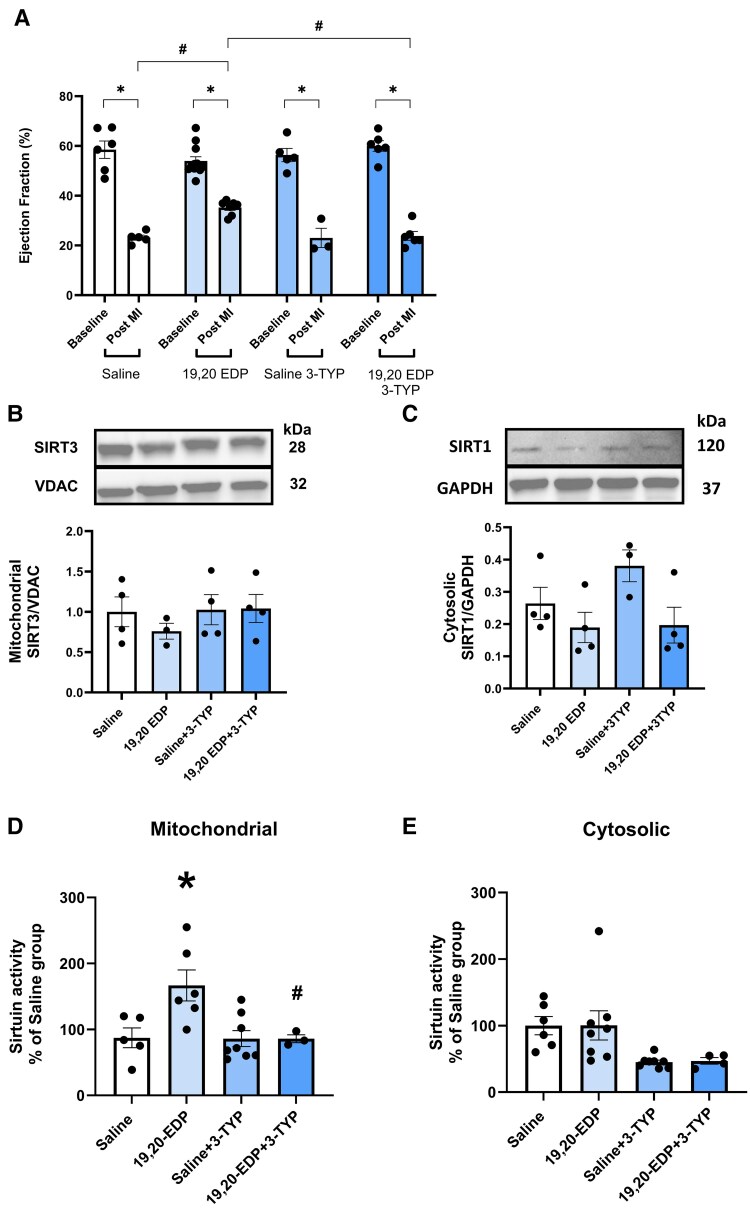
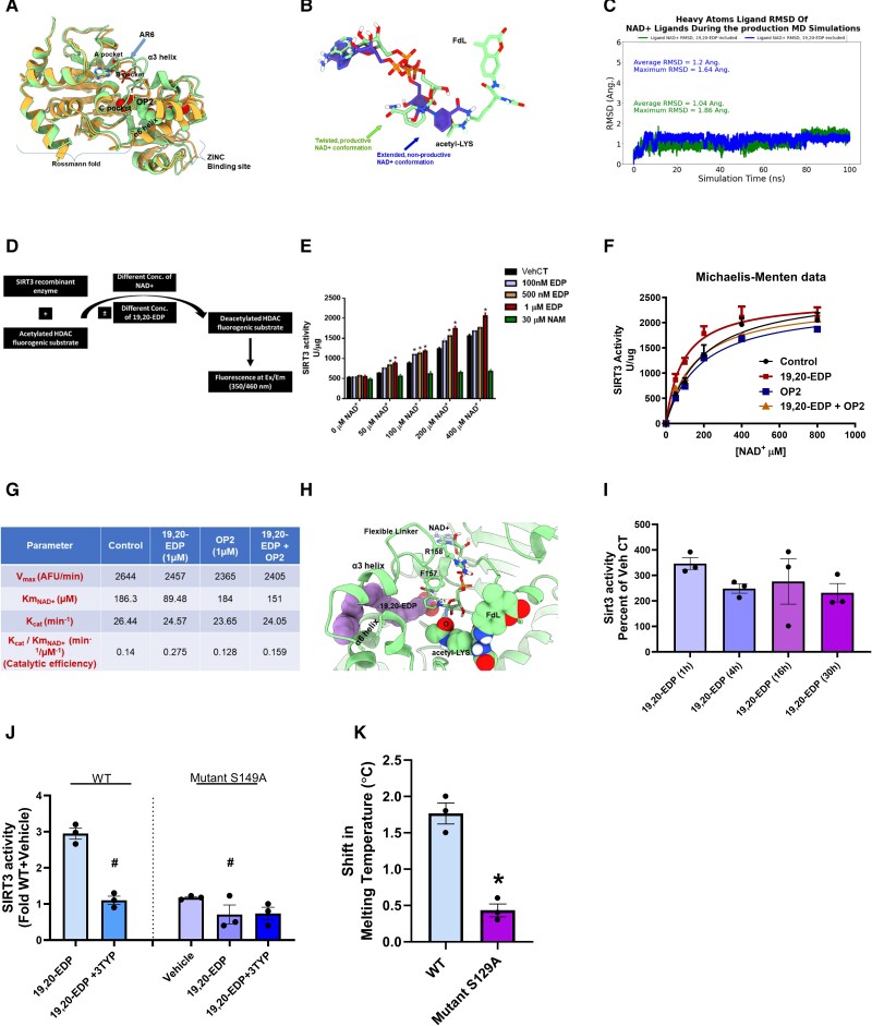
Methods and results: Ex vivo models of isolated heart perfusions were performed in C57BL/6 mice to assess the effect of 19,20-EDP on cardiac function and energy metabolism following IR injury. In vivo permanent occlusion of the left anterior descending coronary artery was performed to induce MI; mice were administered 19,20-EDP with or without the SIRT3 selective inhibitor 3-TYP. Mitochondrial SIRT3 targets and respiration were assessed in human left ventricular tissues obtained from individuals with ischaemic heart disease (IHD) and compared to non-failing controls (NFCs). Binding affinity of 19,20-EDP to human SIRT3 was assessed using molecular modelling and fluorescence thermal shift assay. Results demonstrated that hearts treated with 19,20-EDP had improved post-ischaemic cardiac function, better glucose oxidation rates, and enhanced cardiac efficiency. The cardioprotective effects were associated with enhanced mitochondrial SIRT3 activity. Interestingly, treatment with 19,20-EDP markedly improved mitochondrial respiration and SIRT3 activity in human left ventricle (LV) fibres with IHD compared to NFC. Moreover, 19,20-EDP was found to bind to the human SIRT3 protein enhancing the NAD+-complex stabilization leading to improved SIRT3 activity. Importantly, the beneficial effects of 19,20-EDP were abolished by SIRT3 inhibition or using the S149A mutant SIRT3.

Conclusion: These data demonstrate that 19,20-EDP-mediated cardioprotective mechanisms against ischaemia and IR injury involve mitochondrial SIRT3, resulting in improved cardiac efficiency.

Keywords: 19,20-Epoxydocosapentaenoic acid; Ischaemic human hearts; Ischaemic injury; Mitochondria; Sirtuin 3.


Conflict of interest statement

Conflict of interest: none declared.

Figures

Graphical Abstract
Graphical Abstract
A schematic model illustrating the proposed mechanism of 19,20-EDP in ameliorating cardiac IR injury. SIRT3 activity is suppressed following IR injury resulting in hyperacetylation of several key mitochondrial proteins. This in turn contributes to impaired mitochondrial energy metabolism and function leading to cardiac dysfunction. The current study identifies SIRT3 as a potential target for the epoxylipid 19,20-EDP and reveals that direct binding of 19,20-EDP to SIRT3 significantly enhances its enzymatic activity. 19,20-EDP, via directly activating SIRT3, attenuated IR injury through preventing mitochondrial damage and improving mitochondrial quality as evidenced by enhanced antioxidant capacity, limited oxidative injury, stimulated glucose oxidation, improved mitochondrial respiration, and increased ATP production.
Figure 1
Figure 1
Hearts perfused with 19,20-EDP (10 min before ischaemia) in the ex vivo working heart experiment showed improved post-ischaemic cardiac recovery with higher glucose oxidation rate and cardiac efficiency. (A) Cardiac output, (B) cardiac work, (C) developed pressure, (D) peak systolic pressure, (E) heart rate, (F) rate pressure product, (G) coronary flow, and (H) aortic outflow assessed at the baseline before drug treatment (10 and 20 min), during drug treatment (30 min), during ischaemia, and at 10, 20, 30, and 40 min of reperfusion. (I) Glucose oxidation rate, (J) palmitate (fatty acid) oxidation rate, and (K) cardiac efficiency in hearts subjected to IR injury at the baseline and during reperfusion. Values represent mean ± SEM; significant differences were determined using one-way ANOVA followed by a Tukey post hoc analysis or unpaired, two-tailed Student’s t-test; *P < 0.05 vs. vehicle IR, #P < 0.05 vs. vehicle aerobic (n = 5–8 per group).
Figure 2
Figure 2
Perfusion of mice hearts with 19,20-EDP in the Langendorff mode improved post-ischaemic contractile parameters via maintenance of SIRT3 activity. (A) Representative immunoblot and densitometric quantification of the expression of mitochondrial protein SIRT3 in mice hearts after 30 min ischaemia and 40 min reperfusion. Protein expression was normalized to voltage-dependent anion channel (VDAC) protein used as a loading control. (B) Cardiac mitochondrial sirtuin activity was determined in mitochondrial fractions in mice hearts after 30 min ischaemia and 40 min reperfusion; *P < 0.05 vs. vehicle IR, #P < 0.05 vs. vehicle aerobic. Representative immunoblots and densitometric quantification of the relative protein expression of (C) acetyl MnSOD K68 and (D) acetyl MnSOD K122 normalized to total manganese superoxide dismutase (MnSOD) in mice hearts after 30 min ischaemia and 40 min reperfusion. (E) LVDP recovery at 40 min of reperfusion as a percentage of baseline; *P < 0.05 vs. vehicle IR, #P < 0.05 vs. 19,20-EDP. (F) Representative immunoblot and densitometric quantification of the expression of mitochondrial protein SIRT3 in Sirt3−/− mice hearts after 30 min ischaemia and 40 min reperfusion. (G) Myocardial infarction level is stained by 23,5-triphenyl tetrazolium chloride (TTC) in mice hearts after 30 min ischaemia and 40 min reperfusion. White area indicates infarction (scale bar 50 mm). (H) Myocardial infarct size level normalized to total area. Values represent mean ± SEM; significant differences were determined using one-way ANOVA followed by a Tukey post hoc analysis; *P < 0.05 vs. vehicle IR, #P < 0.05 vs. vehicle aerobic (n = 5–8 per group). LVDP, left ventricular developed pressure.
Figure 3
Figure 3
In vivo administration of 19,20-EDP improved post-MI cardiac function recovery in mice via SIRT3. (A) Left ventricular ejection fraction (EF)% measured by echocardiography. Representative immunoblots and quantification of (B) SIRT3 protein in hearts of mice following MI in mitochondrial fraction and (C) SIRT1 protein in cytosolic fraction. (D) Mitochondrial sirtuin and (E) cytosolic sirtuin activities normalized against saline-treated groups. Values represent mean ± SEM; significant differences were determined using one-way or two-way ANOVA followed by a Tukey post hoc analysis; *P < 0.05 vs. saline treatment only group, #P < 0.05 vs. EDP treatment only group (n = 3–8 per group).
Figure 4
Figure 4
Mitochondrial respiration assessed in permeabilized fresh fibres isolated from mice hearts demonstrated reduced capacity for glucose oxidation when SIRT3 is inhibited. Mitochondrial oxidation of carbohydrates was assessed upon subsequent addition of (A) malate and pyruvate (M + Py), (B) ADP, (C) FCCP, and (D) RCR was calculated as the ratio of mitochondrial respiration in the presence of ADP to mitochondrial respiration in the presence of M + Py. Mitochondrial oxidation of fatty acids was assessed upon subsequent addition of (E) malate and palmitoyl carnitine (M + Pc) (F) ADP, (G) FCCP, and (H) RCR was calculated as the ratio of mitochondrial respiration in the presence of ADP to mitochondrial respiration in the presence of M + Pc. Values represent mean ± SEM; significant differences were determined using one-way ANOVA followed by a Tukey post hoc analysis; *P < 0.05 vs. vehicle, #P < 0.05 vs. EDP CT (n = 5).
Figure 5
Figure 5
Deficiency of 19,20-EDP in human ischaemic left ventricular tissues was associated with reduced SIRT3 expression and activity. (A) 19,20-EDP levels in human left ventricle (LV) tissue (ng/g tissue) were measured by LC–MS/MS. (B) Representative immunoblot and densiometric quantification of the expression of mitochondrial protein SIRT3 and (C) mitochondrial sirtuin activity in non-failing control hearts as well as non-infarct, peri-infarct, and infarct regions of human ischaemic left ventricular tissues. Representative immunoblots and densiometric quantification of the relative protein expression of the mitochondrial (D) AcMnSOD K68, (E) AcMnSOD K122, (F) overall cardiac protein (lysine) acetylation in non-failing control hearts as well as non-infarct, peri-infarct, and infarct regions of human ischaemic left ventricular tissues. SIRT3 and overall cardiac protein (lysine) acetylation were normalized to VDAC, AcMnSOD K68 and AcMnSOD K122 were normalized to total manganese superoxide dismutase (MnSOD). Assessment of mitochondrial sirtuin activity in (G) non-infarct and (H) peri-infarct regions of human ischaemic left ventricular tissues after incubation with the vehicle, 19,20-EDP, NAM, or 19,20-EDP + NAM, data are represented as relative percentage to vehicle treatment. Treatment with 19,20-EDP improved mitochondrial respiratory function in human ischaemic left ventricular tissues. Bar charts demonstrating changes in the values of (I) rate of ATP production in respiration medium in both NFC hearts and non-infarct regions of human ischaemic left ventricular tissues, (J) coupling efficiency, (K) spare respiratory capacity, and (L) respiratory control ratio (RCR). Values represent mean ± SEM; significant differences were determined using one-way ANOVA followed by a Tukey post hoc analysis; &P < 0.05 vs. 19,20-EDP, *P < 0.05 vs. NFC vehicle, #P < 0.05 vs. vehicle non-infarct (n = 3–5 per group).
Figure 6
Figure 6
Treatment of NRCMs, subjected to HR, with 19,20-EDP limited ROS production, preserved mitochondrial density/morphology and maintained mitochondrial membrane potential. Representative images of NRCMs stained with either (MitoSOX and Hoechst 33342 dyes), or (MitoTracker green, TMRE, and Hoechst 33342 dyes) (scale bar 5 um). Histograms representing the (A) the quantification of percentage of MitoSOX fluorescence intensity, (B) quantification of percentage of NRCMs with preserved mitochondrial mass as evidenced by MitoTracker stain, (C) the relative quantification of TMRE fluorescence intensity. Normal mitochondrial morphology (filamentous and tubular shape) is highlighted by arrows shown in cells from aerobic control and 19,20-EDP treatments and cells treated with 19,20-EDP under hypoxia/reoxygenation. In contrast, punctate and fragmented mitochondrial morphology are highlighted by the other arrows. Values represent mean ± SEM; significant differences were determined using one-way ANOVA followed by a Tukey post hoc analysis; *P < 0.05 vs. aerobic vehicle, #P < 0.05 vs. HR vehicle (n = 3 per group, five fields each).
Figure 7
Figure 7
19,20-EDP directly binds and enhances the activity of the mitochondrial SIRT3. (A) A structure superposition of the 4BN4 structure (light-green cartoon representation) as a representative for structures with an open allosteric pocket over the crystal structures of 5Z93 (orange cartoon representation) as a representative of structures with closed pocket. The 4BN4 structure is co-crystallized with AR6 (a NAD+ analogue without the nicotinamide motif), OP2 (shown as vdW spheres, with light-green carbon and red oxygen atoms) that binds the allosteric pocket and whose binding requires an apparent clockwise motion of the α3 helix. (B) A superposition for the three-dimensional structures of NAD+ in the twisted productive conformation overlaid on the extended non-productive conformation. The extended conformation exhibits steric clashes with the acetyl-LYS binding site of the modelled FdL dye. (C) Ligand heavy atoms RMSD plot of NAD+ in the presence and in the absence of 19,20-EDP during the 100 ns production MD simulations. (D) A schematic diagram illustrating the experiment used for the assessment of the direct effect of 19,20-EDP on the activity SIRT3. (E) Bar chart showing the effect of different concentrations of 19,20-EDP on the activity of SIRT3 in the presence of different concentrations of the cofactor NAD+. (F) Michaelis–Menten fit for SIRT3 and its cofactor NAD+ in the presence of the vehicle, 19,20-EDP (1 µM), OP2 (1 µM), or 19,20-EDP (1 µM) + OP2 (1 µM). (G) SIRT3 kinetic values obtained using a Michaelis–Menten fit in the presence of the vehicle, 19,20-EDP, OP2, or 19,20-EDP + OP2. (H) A model structure of the full reaction-ready complex of hSIRT3 (shown in light-green cartoons) with bound allosteric modulator (19,20-EDP, shown as vdW spheres, with purple carbons and red oxygen atoms), cofactor (NAD+, shown in stick representation with light-green 25 carbon, orange phosphorous, red oxygen, blue nitrogen, and white hydrogen atoms) and a model substrate (FdL, shown as vdW spheres with light-green carbons, red oxygen, blue nitrogen, and white hydrogen atoms). The side chains of critical flexible linker residues those are important for NAD+ binding (PHE157 and ARG158) are shown in ball and stick representations, with light-green carbon, yellow nitrogen, and white hydrogen atoms. The first step of the proposed catalysis is the nucleophilic attack of the acetyl-oxygen of the substrate on the electron deficient anomeric C1`` atom of the NAD+ molecule. (I) Bar chart demonstrating the time-dependent effect of 19,20-EDP (1 µM) on SIRT3 activity in NRCMs. (J) Wild-type (WT) or S149A mutant SIRT3 was assessed for deacetylase activity in a cell-free fluorogenic assay at NAD+-depleted conditions (50 µM) in the absence or presence of 1 µM 19,20-EDP with/without SIRT3 inhibitor 3-TYP. (K) Binding affinity was assessed between human recombinant SIRT3 protein (WT or mutant S129A) and 19,20-EDP using a fluorescence based thermal shift assay. Data are represented as mean ± SEM. Significant differences were determined using one-way ANOVA followed by a Tukey post hoc analysis; *P < 0.05 vs. vehicle, #P < 0.05 vs. WT 19,20-EDP (n = 3). Or one sample t-test comparing melting temperature of SIRT3 WT vs. mutant.

References

    1. Martin SS, Aday AW, Almarzooq ZI, Anderson CAM, Arora P, Avery CL, Baker-Smith CM, Gibbs BB, Beaton AZ, Boehme AK, Commodore-Mensah Y, Currie ME, Elkind MSV, Evenson KR, Generoso G, Heard DG, Hiremath S, Johansen MC, Kalani R, Kazi DS, Ko D, Liu J, Magnani JW, Michos ED, Mussolino ME, Navaneethan SD, Parikh NI, Perman SM, Poudel R, Rezk-Hanna M, Roth GA, Shah NS, St-Onge M-P, Thacker EL, Tsao CW, Urbut SM, Spall HGCV, Voeks JH, Wang N-Y, Wong ND, Wong SS, Yaffe K, Palaniappan LP. 2024 heart disease and stroke statistics: a report of US and global data from the American Heart Association. Circulation 2024;149:e347–e913. - PubMed
    1. Hausenloy DJ, Yellon DM. Myocardial ischemia-reperfusion injury: a neglected therapeutic target. J Clin Invest 2013;123:92–100. - PMC - PubMed
    1. Perrelli M-G, Pagliaro P, Penna C. Ischemia/reperfusion injury and cardioprotective mechanisms: role of mitochondria and reactive oxygen species. World J Cardiol 2011;3:186. - PMC - PubMed
    1. Michishita E, Park JY, Burneskis JM, Barrett JC, Horikawa I. Evolutionarily conserved and nonconserved cellular localizations and functions of human SIRT proteins. Mol Biol Cell 2005;16:4623–4635. - PMC - PubMed
    1. Rardin MJ, Newman JC, Held JM, Cusack MP, Sorensen DJ, Li B, Schilling B, Mooney SD, Kahn CR, Verdin E. Label-free quantitative proteomics of the lysine acetylome in mitochondria identifies substrates of SIRT3 in metabolic pathways. Proc Natl Acad Sci U S A 2013;110:6601–6606. - PMC - PubMed

MeSH terms

LinkOut - more resources