Review
. 2025 Jun;292(12):2992-3013.
doi: 10.1111/febs.17356. Epub 2024 Dec 10.

Protein import into bacterial endosymbionts and evolving organelles

Affiliations
Review

Protein import into bacterial endosymbionts and evolving organelles

Megan E S Sørensen et al. FEBS J. 2025 Jun.

Abstract

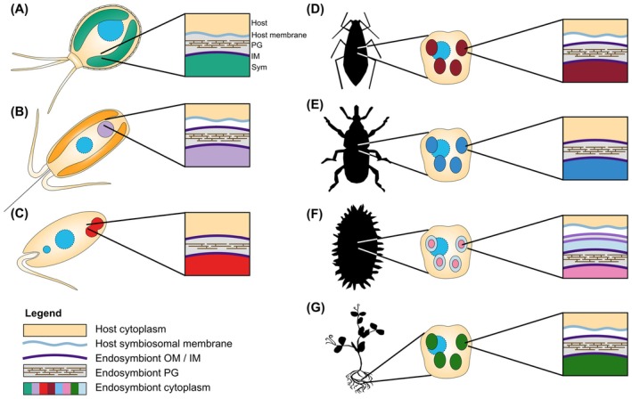
Bacterial endosymbionts are common throughout the eukaryotic tree of life and provide a range of essential functions. The intricate integration of bacterial endosymbionts into a host led to the formation of the energy-converting organelles, mitochondria and plastids, that have shaped eukaryotic evolution. Protein import from the host has been regarded as one of the distinguishing features of organelles as compared to endosymbionts. In recent years, research has delved deeper into a diverse range of endosymbioses and discovered evidence for 'exceptional' instances of protein import outside of the canonical organelles. Here we review the current evidence for protein import into bacterial endosymbionts. We cover both 'recently evolved' organelles, where there is evidence for hundreds of imported proteins, and endosymbiotic systems where currently only single protein import candidates are described. We discuss the challenges of establishing protein import machineries and the diversity of mechanisms that have independently evolved to solve them. Understanding these systems and the different independent mechanisms, they have evolved is critical to elucidate how cellular integration arises and deepens at the endosymbiont to organelle interface. We finish by suggesting approaches that could be used in the future to address the open questions. Overall, we believe that the evidence now suggests that protein import into bacterial endosymbionts is more common than generally realized, and thus that there is an increasing number of partnerships that blur the distinction between endosymbiont and organelle.

Keywords: Paulinella; Strigomonadinae; UCYN‐A; endosymbiosis; envelope membranes; mealybug; nitroplast; organellogenesis; protein translocation; targeting signals.


Conflict of interest statement

The authors declare no conflict of interest.

Figures

Fig. 1
Fig. 1
Synchronized cell division of endosymbiont‐harboring protists with their endosymbionts. (A) Paulinella chromatophora, (B) Braarudosphaera bigelowii, (C) Angomonas deanei. C, chromatophore; K, kinetoplast (a network of circular DNA containing many copies of the mitochondrial genome typical for the kinetoplastida); N, nucleus; P, plastid; S, endosymbiont.
Fig. 2
Fig. 2
Capacity of endosymbionts to autonomously build a bacterial cell envelope. (A) Gene presence/absence patterns in the genomes of endosymbiotic (red) and free‐living bacteria (black) as analyzed by the toolset provided by the Kyoto Encyclopedia for Genes and Genomes (https://www.kegg.jp/). (B–E) As a reference, pathways for phospholipid biosynthesis (B), PG biosynthesis (C), LPS biosynthesis (D), and the envelope structure of Escherichia coli (E) are provided.
Fig. 3
Fig. 3
Visualizations of the endosymbiotic systems, highlighting the membrane systems surrounding the endosymbionts that any imported proteins need to traverse. (A) Paulinella chromatophora and its chromatophores. (B) Braarudosphaera bigelowii and UCYN‐A, nitroplast. (C) Angomonas deanei, as a representative of the Strigomonadinae, and Ca. Kinetoplastibacterium (D) Pea aphid, Acyrthosiphon pisum, and Buchnera endosymbiont shown within a bacteriocyte. (E) Representing both the red palm weevil, Rhynchophorus ferrugineus, and its Nardonella endosymbiont, and the cereal weevil, Sitophilus, and its endosymbiont Sodalis pierantonius. Shown within a bacteriocyte. (F) Mealybug, Planococcus citri, and its nested endosymbionts, Ca. Tremblaya princeps and Ca. Moranella endobia, shown within a bacteriocyte. (G) A legume and its rhizobia endosymbionts shown within a root nodule cell.
Fig. 4
Fig. 4
Comparison of endosymbiotic‐targeted proteins. (A) Chromatophore‐targeted proteins (CTP) in Paulinella chromatophora form two groups, short (< 90 aa) and long (> 250 aa) CTPs. (B) Schematic alignment of 3 lCTPs showing the characteristic bipartite N‐terminal extension (crTP, green). The zoom‐in shows a multiple sequence alignment of 12 representative crTPs. (C) Schematic alignment of 3 nitroplast‐targeted proteins (NTP) showing the characteristic C‐terminal extension (uTP, red). The zoom‐in shows a multiple alignment of 12 representative uTPs. Scissors symbols mark the targeting signal cleavage sites deduced by MS analyses in B and C. (D) Multiple sequence alignments of 7 BCRs from the aphid Acyrthosiphon pisum and NCRs from the legume Medicago truncatula. Six representative NCRs of group A (4 conserved cysteines) and group B (6 conserved cysteines) are shown. Signal peptides are underlined, and asterisks show conserved cysteine residues. For all alignments, amino acids are color‐coded in blue (positively charged; H, K, R), magenta (negatively charged; E, D), cyan (hydrophobic; A, P, W, I, L, M, V, F), orange (neutral; G, S, Y, N, Q, T), and black (cysteines; C). Sequence identifiers or accession numbers are, from top to bottom, for crTPs: scaffold7571‐size1527|m.60359; scaffold3807‐size2095|m.37686; scaffold10361‐size1249|m.74090; scaffold6875‐size1609|m.56608; scaffold8035‐size1476|m.62771; scaffold3865‐size2079|m.38081; scaffold5513‐size1797|m.48594; scaffold4638‐size1933|m.43151; scaffold2991‐size2309|m.31974; scaffold4337‐size1989|m.41170; scaffold2706‐size2392|m.29779; scaffold2155‐size2616|m.25388 [see ref. ; available at PRIDE Repository (https://www.ebi.ac.uk/pride/archive/), accession number PXD006531]; for uTPs: KC1‐P2‐N_CL7753Contig1_1; KC1‐P2‐N_CL1062Contig1_1; KC1‐P2‐N_CL4024Contig1_1; KC1‐P2‐N_CL8449Contig1_1; KC1‐P2‐N_CL1190Contig1_1; KC1‐P2‐N_CL2249Contig1_1; KC1‐P2‐N_CL4289Contig1_1; KC1‐P2‐N_CL7819Contig1_1; KC1‐P2‐N_CL7868Contig1_1; KC1‐P2‐N_CL1296Contig1_1; KC1‐P2‐N_CL2661Contig1_1; KC1‐P2‐N_k25_Locus_10184_Trans [see ref. , available at Dryad, https://doi.org/10.5061/dryad.2z34tmptf]; for BCRs (AphidBase IDs): ACYPI32128; ACYPI38738; ACYPI44142; AK343177; AK339855; ACYPI49532; ACYPI45157; for NCRs (NCBI accession no.) group a: AFK48426.1; AES68835.1; ABS31393.1; RHN51124.1; AES98754.1; ABS31399.1 and group b: ABS31396.1; ABS31401.1; KEH26626.1; XP_039683422.1; KEH38199.1; AES78310.1. Sequence alignments were made with clustalx (B, C) or clustalw (D) and refined manually.

Similar articles

Cited by

References

    1. Martin W, Rujan T, Richly E, Hansen A, Cornelsen S, Lins T, Leister D, Stoebe B, Hasegawa M & Penny D (2002) Evolutionary analysis of Arabidopsis, cyanobacterial, and chloroplast genomes reveals plastid phylogeny and thousands of cyanobacterial genes in the nucleus. Proc Natl Acad Sci USA 99, 12246–12251. - PMC - PubMed
    1. Timmis JN, Ayliffe MA, Huang CY & Martin W (2004) Endosymbiotic gene transfer: organelle genomes forge eukaryotic chromosomes. Nat Rev Genet 5, 123–135. - PubMed
    1. Nowack ECM & Weber APM (2018) Genomics‐informed insights into endosymbiotic organelle evolution in photosynthetic eukaryotes. Annu Rev Plant Biol 69, 51–84. - PubMed
    1. Roger AJ, Muñoz‐Gómez SA & Kamikawa R (2017) The origin and diversification of mitochondria. Curr Biol 27, R1177–R1192. - PubMed
    1. Chen LJ & Li HM (2017) Stable megadalton TOC–TIC supercomplexes as major mediators of protein import into chloroplasts. Plant J 92, 178–188. - PubMed

Substances

Associated data

LinkOut - more resources