

Selective PET imaging of CXCR4 using the Al18F-labeled antagonist LY2510924
- PMID: 39658737
- PMCID: PMC11928405
- DOI: 10.1007/s00259-024-07025-w
Selective PET imaging of CXCR4 using the Al18F-labeled antagonist LY2510924
Abstract
Background: [68Ga]PentixaFor detects C-X-C chemokine receptor type 4 (CXCR4) overexpression in various malignancies, such as multiple myeloma and non-Hodgkin lymphomas, as well as in endocrine and inflammatory disorders. This study aimed to develop an Al18F-labeled radiotracer derived from LY2510924 for CXCR4-targeted imaging, leveraging the physical and logistical advantages of fluorine-18.
Methods: We designed a CXCR4-specific radioprobe, [18F]AlF-NOTA-SC, based on LY2510924 by incorporating a triglutamate linker and NOTA chelator to enable Al18F-labeling. The in vitro CXCR4 affinity was assessed using cell-based binding assays. Subsequently, in vivo pharmacokinetics and tumor uptake of [18F]AlF-NOTA-SC were assessed in naïve mice and mice with xenografts derived from U87.CD4/U87.CD4.CXCR4 and MM.1 S cells. Finally, biodistribution was determined in a non-human primate using PET-MR.
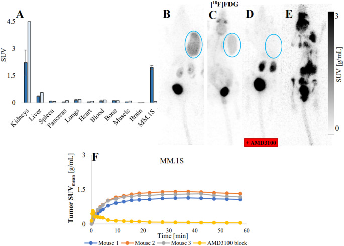
Results: Compared to Ga-PentixaFor, AlF-NOTA-SC demonstrated similar in vitro affinity for human CXCR4. [18F]AlF-NOTA-SC was produced with a decay-corrected radiochemical yield of 21.0 ± 7.1% and an apparent molar activity of 16.4 ± 3.6 GBq/µmol. In [18F]AlF-NOTA-SC binding assays on U87.CD4.CXCR4 cells, the total bound fraction was 7.1 ± 0.5% (58% blocking by AMD3100). In naïve mice, the radiotracer did not accumulate in any organs; however, it showed a significant CXCR4-specific uptake in xenografted tumors (SUVmeanU87.CD4 = 0.04 ± 0.00 (n = 3); SUVmeanU87.CD4.CXCR4 = 3.04 ± 0.65 (n = 3); SUVmeanMM.1 S = 1.95 ± 0.11 (n = 3)). In a non-human primate, [18F]AlF-NOTA-SC accumulated in CXCR4 expressing organs, such as the spleen and bone marrow.
Conclusion: [18F]AlF-NOTA-SC exhibited CXCR4-specific uptake in vitro and in vivo, with fast and persistent tumor accumulation, making it a strong candidate for clinical translation as an 18F-alternative to [68Ga]PentixaFor.
Keywords: Al18F; Al18F-NOTA-SC; CXCR4; PET.
© 2024. The Author(s).
Conflict of interest statement
Declarations. Research involving human participants and/or animals: All of the herein discussed animal studies were conducted according to the international, national, and institutional regulations governing the care and use of animals. Each experiment was approved by the KU Leuven ethical review board (Reference P010/2023 (mice studies) and P112/2019 (non-human primate study)). This research did not involve any human material or human participants. Informed consent: Not applicable. Consent for publication: All the authors approved the publication of the article. Conflict of interest: The authors have no relevant financial or non-financial interests to disclose.
Figures


References
-
- Balkwill F. The significance of cancer cell expression of the chemokine receptor CXCR4., Semin. Cancer Biol., Jun. 2004;14(3):171–179.10.1016/j.semcancer.2003.10.003 - PubMed
-
- Domanska UM et al. A review on CXCR4/CXCL12 axis in oncology: no place to hide., Eur. J. Cancer Oxf. Engl. 1990, Jan. 2013;49(1):219–230.10.1016/j.ejca.2012.05.005 - PubMed
-
- Guo F, Wang Y, Liu J, Mok SC, Xue F, Zhang W. CXCL12/CXCR4: a symbiotic bridge linking cancer cells and their stromal neighbors in oncogenic communication networks. Oncogene. Feb. 2016;35(7):816–26. 10.1038/onc.2015.139. - PubMed
MeSH terms
Substances
Grants and funding
LinkOut - more resources
Full Text Sources
Research Materials
