

Successful diagnosis and treatment of canine polymyositis: utilizing MRI and immunohistochemistry for accurate detection
- PMID: 39658799
- PMCID: PMC11629501
- DOI: 10.1186/s12917-024-04356-6
Successful diagnosis and treatment of canine polymyositis: utilizing MRI and immunohistochemistry for accurate detection
Abstract
Background: Inflammatory myopathy is generally categorized into generalized inflammatory myopathies (gIM), which affect muscles throughout the body, and focal inflammatory myopathies (fIM), which are localized to specific muscles or muscle groups. This report details a case of immune-mediated polymyositis in a dog, successfully diagnosed using MRI and IHC and managed with immunosuppressive therapy.
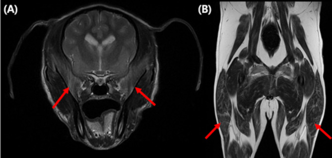
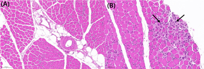
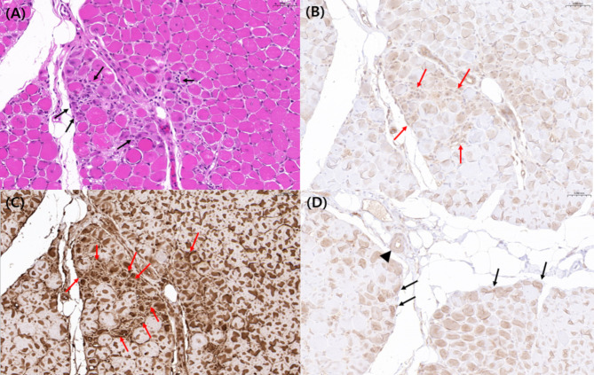
Case presentation: A 5-year-old castrated male Poodle was admitted to our hospital presenting with lethargy and exercise intolerance. Biochemical analysis revealed significantly elevated serum levels of aspartate aminotransferase (AST) and creatine kinase (CK). Physical examination showed muscle atrophy in the hind legs, but further orthopedic and neurological examinations identified no additional abnormalities. MRI demonstrated hyperintense and heterogeneous signal changes across the muscles, including contrast enhancement, suggesting inflammatory myopathy. This diagnosis was confirmed through histopathological examination, which revealed inflammatory lesions with fibrous tissue proliferation within the muscle tissue. To investigate the presence and type of inflammatory cells and vascular changes, aiding in the differential diagnosis of inflammatory myopathies, immunohistochemistry (IHC) was performed, revealing positive findings for CD8+, CD4+, and VEGF in the evaluated tissue, leading to a diagnosis of polymyositis.
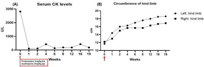
Conclusions: The dog was diagnosed with immune-mediated polymyositis and treatment was initiated with prednisolone at 1 mg/kg twice daily and azathioprine at 2 mg/kg once daily. Following the administration of these immunosuppressive agents, CK levels returned to normal, and the dog's exercise intolerance and lethargy resolved. The thickness of the hind legs also increased progressively. The dog has maintained an improved condition under continued immunosuppressive therapy for four months. This case highlights the critical role of MRI and immunohistochemistry in diagnosing immune-mediated polymyositis, demonstrating their alternative capability in cases where conventional electromyography (EMG) is not feasible in this context.
Keywords: Dog; Immune-mediated; MRI; Polymyositis.
© 2024. The Author(s).
Conflict of interest statement
Declarations. Ethics approval and consent to participate: Not applicable. Consent for publication: Written informed consent was obtained from the dog’s owner. Competing interests: The authors declare no competing interests.
Figures
References
-
- Evans J, Levesque D, Shelton GD. <ArticleTitle Language=“En”>Canine inflammatory myopathies: a clinicopathologic review of 200 cases. J Vet Intern Med. 2004;18(5):679–91. - PubMed
-
- Dalakas MC, Hohlfeld R. Polymyositis and dermatomyositis. Lancet. 2003;362(9388):971–82. - PubMed
-
- Shelton GD. From dog to man: the broad spectrum of inflammatory myopathies. Neuromuscul Disord. 2007;17(9–10):663–70. - PubMed
-
- Kornegay JN, Gorgacz EJ, Dawe DL, Bowen JM, White NA, et al. Polymyositis in dogs. J Am Vet Med Assoc. 1980;176(5):431–8. - PubMed
-
- MOROZUMI M, OYAMA Y, KUROSU Y, NAKAYAMA H, GOTO N, et al. Immune-mediated polymyositis in a dog. J Vet Med Sci. 1991;53(3):511–2. - PubMed
Publication types
MeSH terms
Substances
Grants and funding
LinkOut - more resources
Full Text Sources
Medical
Research Materials
