

Curve Progression After the Termination of Bracing for Adolescent Idiopathic Scoliosis: Usefulness of Combining the Proximal Femur Maturity Index (PFMI) and Risser Staging
- PMID: 39659310
- PMCID: PMC11631167
- DOI: 10.7759/cureus.73395
Curve Progression After the Termination of Bracing for Adolescent Idiopathic Scoliosis: Usefulness of Combining the Proximal Femur Maturity Index (PFMI) and Risser Staging
Abstract
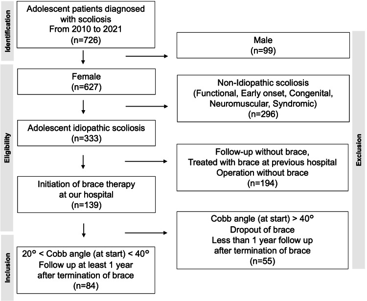
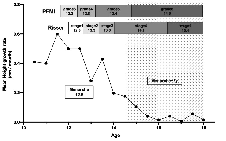
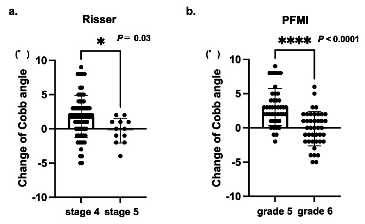
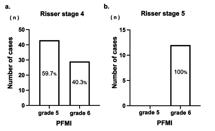
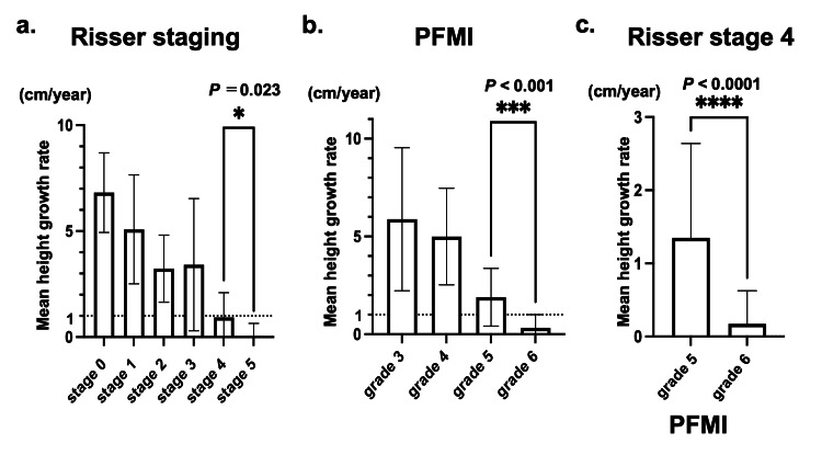
Background The brace therapy for adolescent idiopathic scoliosis (AIS) typically ends upon the end of growth. However, determining the timing of growth cessation can be challenging. The purpose of this study was to evaluate the utility of the proximal femur maturity index (PFMI), which can be assessed simultaneously with Risser staging without requiring additional radiation exposure, in determining the appropriate timing to terminate bracing. To achieve this, we investigated the relationship between skeletal maturity at the end of bracing, post-bracing curve progression, and height growth in patients who had been successfully treated with a brace. Methods Between April 2010 and March 2021, a total of 84 female patients with AIS who started bracing at our hospital with an initial Cobb angle of 20-40 degrees were included. All patients were followed for at least one year after brace termination. Height and radiographic parameters (Risser staging, PFMI, Cobb angle) were retrospectively collected. Results At the end of the bracing period, patients were categorized into Risser stage 4 (85.7%) and 5 (14.3%). By the last follow-up, patients with Risser stage 4 experienced an average main curve progression of 1.8°, whereas those with Risser stage 5 had an average progression of -0.3° (P = 0.03). Patients with Risser stage 4 were further divided into PFMI grade 5 (59.7%) and 6 (40.3%). Significant curve progression was observed in patients with PFMI grade 5 (average: 3.0°) compared to grade 6 (average: -0.6°) (P < 0.0001). The mean height growth was 1.9 cm/year for PFMI grade 5, and 0.3 cm/year for grade 6, with significant differences between these groups (P < 0.001). Conclusions PFMI allowed further categorization within Risser stage 4: PFMI grade 5 indicated remaining growth potential and risk of postbracing curve progression, whereas grade 6 indicated growth cessation. The combined use of Risser staging and PFMI, both evaluable through the same whole-spine radiograph, may provide a more accurate prediction of growth cessation.
Keywords: adolescent idiopathic scoliosis (ais); height growth; proximal femur maturity index; risser staging; skeletal maturation.
Copyright © 2024, Shitozawa et al.
Conflict of interest statement
Human subjects: Consent for treatment and open access publication was obtained or waived by all participants in this study. Ethics Committee of Okayama University issued approval No. 2212-021. Animal subjects: All authors have confirmed that this study did not involve animal subjects or tissue. Conflicts of interest: In compliance with the ICMJE uniform disclosure form, all authors declare the following: Payment/services info: All authors have declared that no financial support was received from any organization for the submitted work. Financial relationships: All authors have declared that they have no financial relationships at present or within the previous three years with any organizations that might have an interest in the submitted work. Other relationships: All authors have declared that there are no other relationships or activities that could appear to have influenced the submitted work.
Figures
Similar articles
-
Using the Proximal Femur Maturity Index at Brace Initiation for Adolescent Idiopathic Scoliosis Predicts Curve Progression Risk.J Bone Joint Surg Am. 2024 Mar 20;106(6):531-541. doi: 10.2106/JBJS.23.00694. Epub 2024 Jan 23. J Bone Joint Surg Am. 2024. PMID: 38261654 Free PMC article.
-
Does the Use of Sanders Staging and Distal Radius and Ulna Classification Avoid Mismatches in Growth Assessment with Risser Staging Alone?Clin Orthop Relat Res. 2021 Nov 1;479(11):2516-2530. doi: 10.1097/CORR.0000000000001817. Clin Orthop Relat Res. 2021. PMID: 34036944 Free PMC article.
-
The Utility of a Novel Proximal Femur Maturity Index for Staging Skeletal Growth in Patients with Idiopathic Scoliosis.J Bone Joint Surg Am. 2022 Apr 6;104(7):630-640. doi: 10.2106/JBJS.21.00747. Epub 2022 Jan 6. J Bone Joint Surg Am. 2022. PMID: 35006096
-
Discontinuation of brace treatment in adolescent idiopathic scoliosis (AIS): a scoping review.Spine Deform. 2024 Sep;12(5):1217-1228. doi: 10.1007/s43390-024-00882-3. Epub 2024 May 1. Spine Deform. 2024. PMID: 38693334 Free PMC article.
-
Standardization of criteria for adolescent idiopathic scoliosis brace studies: SRS Committee on Bracing and Nonoperative Management.Spine (Phila Pa 1976). 2005 Sep 15;30(18):2068-75; discussion 2076-7. doi: 10.1097/01.brs.0000178819.90239.d0. Spine (Phila Pa 1976). 2005. PMID: 16166897 Review.
References
-
- Treatment of bracing for adolescent idiopathic scoliosis patients: a meta-analysis. Zhang Y, Li X. Eur Spine J. 2019;28:2012–2019. - PubMed
-
- Maturity assessment and curve progression in girls with idiopathic scoliosis. Sanders JO, Browne RH, McConnell SJ, Margraf SA, Cooney TE, Finegold DN. J Bone Joint Surg Am. 2007;89:64–73. - PubMed
LinkOut - more resources
Full Text Sources
Miscellaneous