

Radicular Dentin Thickness and Root Canal Morphology of Mandibular Incisors in Indian Subpopulation Using Cone Beam Computed Tomography
- PMID: 39659315
- PMCID: PMC11628873
- DOI: 10.7759/cureus.73355
Radicular Dentin Thickness and Root Canal Morphology of Mandibular Incisors in Indian Subpopulation Using Cone Beam Computed Tomography
Abstract
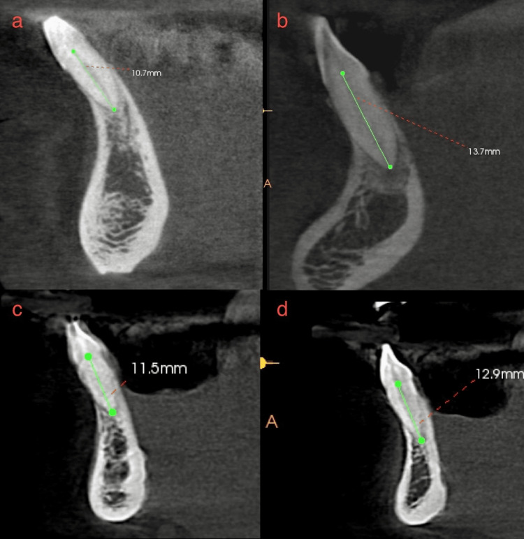
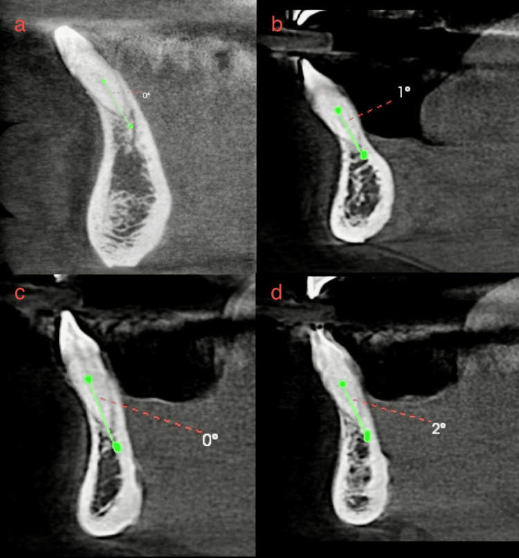
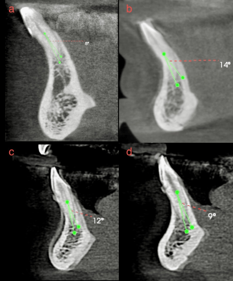
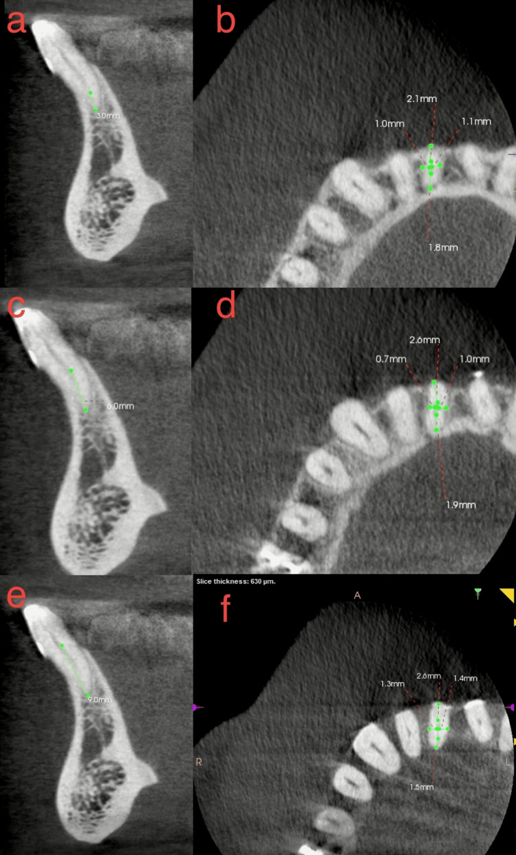
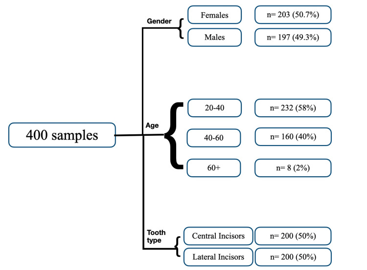
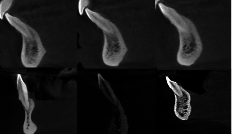
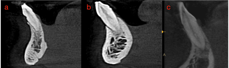
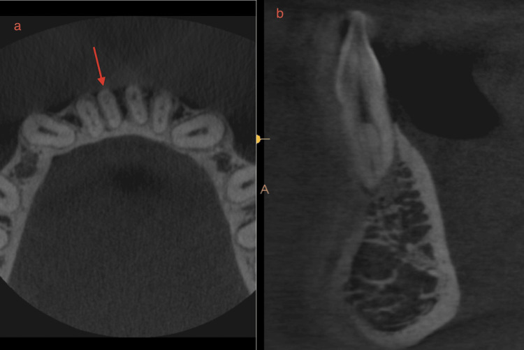
Aim Using two classifications, this study assessed root morphology and canal configuration and measured the Dentin thickness (DT) and canal shapes. Methods Cone beam computed tomography (CBCT) with 400 Mandibular Incisors was collected and assessed for the number, length, curvature of roots, number of canals, bifurcation level, configurations based on Vertucci's and Ahmed's classification, DT and canal shape at 3, 6, 9 mm from the apex. The collected data was subjected to statistical analysis with a level of significance at p<0.05. Results All samples had one root, averaging 12.769 ± 1.128 mm in central incisor (CI) and 13.044 ± 1.235 mm in lateral incisor (LI), with most roots being straight. Most samples had one canal in both teeth, with bifurcations most frequent in the middle third. The most frequent configuration was type 1 Vertucci or 1CI1/1LI1 by Ahmed, followed by type 3 or 1CI1-2-1/1LI1-2-1. One sample, not classifiable under Vertucci, was classified as 1CI1-3-1 by Ahmed. The mean DT for CI was 3.18 ± 0.639 mm, 3.72 ± 0.671 mm and 4.43 ± 0.754 mm labiolingually and 1.578 ± 0.342 mm, 1.881 ± 0.374 mm, 2.283 ± 0.465 mm mesioditally at 3, 6, 9 mm from the apex, respectively. For LI, mean DT was 3.41 ± 0.916 mm, 3.90 ± 0.702 mm and 4.55 ± 0.746 mm labiolingually and 1.63 ± 0.322 mm, 1.981 ± 0.485 mm, 2.55 ± 0.470 mm mesioditally at 3, 6, 9 mm from the apex respectively, canal shape changed from oval to round, from apical to coronal. Conclusion Single canals were the most common, followed by two canals. The middle third of the canal had the most bifurcations. Vertucci type 1 or Ahmed's 1CI1/1LI1 was the most commonly reported canal configuration, with one sample that could not be classified under Vertucci but could be classified using Ahmed classification. DT increased apical to coronal. The canal shape changed from oval to rounded, from apical to coronal.
Keywords: ahmed classification; cone-beam computed tomography (cbct); mandibular incisors; radicular dentin thickness; root canal morphology; vertucci classification.
Copyright © 2024, Krishnan et al.
Conflict of interest statement
Human subjects: Consent for treatment and open access publication was obtained or waived by all participants in this study. Kempegowda Institute of Medical Sciences issued approval KIMS/IEC/D022/D/2022. Animal subjects: All authors have confirmed that this study did not involve animal subjects or tissue. Conflicts of interest: In compliance with the ICMJE uniform disclosure form, all authors declare the following: Payment/services info: All authors have declared that no financial support was received from any organization for the submitted work. Financial relationships: All authors have declared that they have no financial relationships at present or within the previous three years with any organizations that might have an interest in the submitted work. Other relationships: All authors have declared that there are no other relationships or activities that could appear to have influenced the submitted work.
Figures
References
-
- A new system for classifying root and root canal morphology. Ahmed HM, Versiani MA, De-Deus G, Dummer PM. Int Endod J. 2017;50:761–770. - PubMed
-
- Root canal morphology of the permanent mandibular incisors by cone beam computed tomography: a systematic review. Herrero-Hernández Herrero-Hernández, S.; López-Valverde, et al. Appl Sci. 2020;10:4914.
-
- Canal configuration in the mesiobuccal root of the maxillary first molar and its endodontic significance. Weine FS, Healey HJ, Gerstein H, Evanson L. Oral Surg Oral Med Oral Pathol. 1969;28:419–425. - PubMed
LinkOut - more resources
Full Text Sources
Miscellaneous