. 2024 Nov 26:11:1417985.
doi: 10.3389/fmed.2024.1417985. eCollection 2024.

Terbinafine in acrylic polymer for the treatment of onychomycosis in hemodialysis patients: a phase II clinical trial

Affiliations

Terbinafine in acrylic polymer for the treatment of onychomycosis in hemodialysis patients: a phase II clinical trial

Jeanne Marie Queiroz Borges Bersano et al. Front Med (Lausanne). .

Abstract

Introduction: Onychomycosis is a nail infection caused by dermatophyte fungi, non-dermatophyte fungi, and yeast. Patients with chronic kidney disease on dialysis are part of the population that presents higher rates of this disease, mainly due to immunosuppression. Among patients with chronic kidney disease on dialysis, the treatment of onychomycosis is complex, mainly due to the limitations imposed by comorbidities. In this context, the study evaluated the safety and potential efficacy of a treatment that combines nail debridement with the use of acrylic gel nails carrying terbinafine at a concentration of 2%.

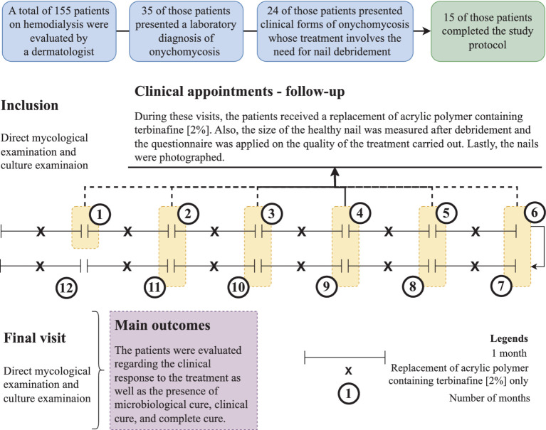
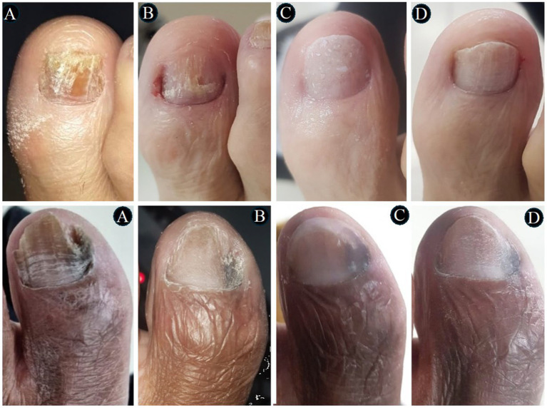
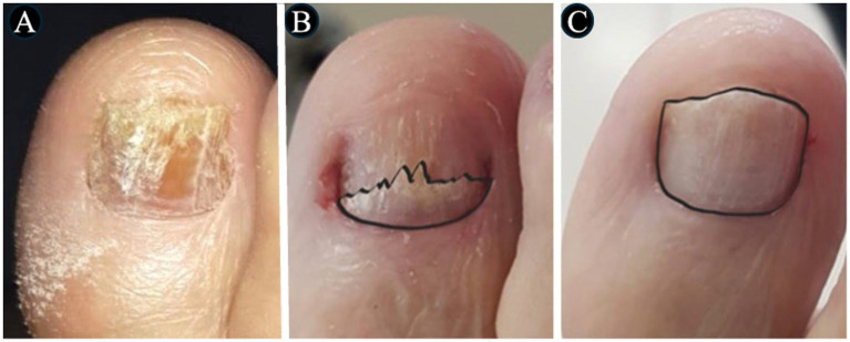
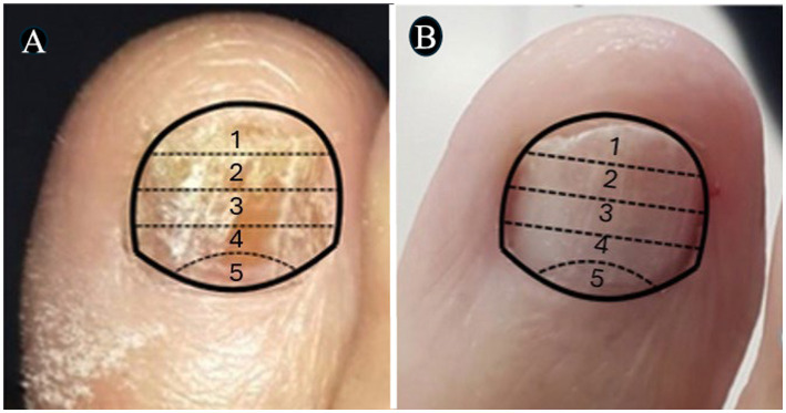
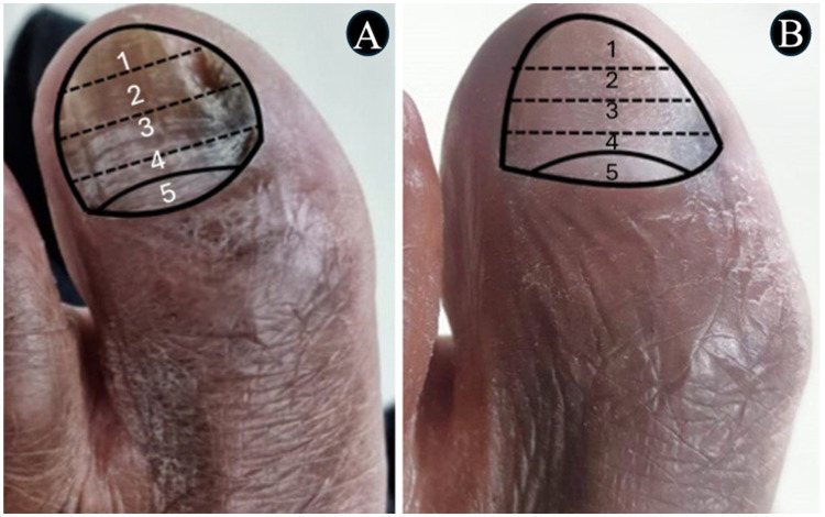
Methods: Patients from the Hemodialysis Center of the São Francisco de Assis University Hospital in Bragança Paulista, São Paulo, Brazil were included. Those had hallux onychomycosis with clinical forms whose treatment involved the need for nail debridement. After the debridement procedure, a nail prosthesis made with acrylic reconstruction gel and 2% terbinafine was applied. The procedure was renewed every 2 weeks (~14 days) for 11 months. The evolution was monitored with measurements of the normal-appearing nail plate and photographs. Direct mycological examination and fungal culture were performed at the beginning of the study and 30 days after applications were interrupted. Assessment of clinical response, clinical cure, mycological cure, and complete cure was performed at the end of the study. All participants answered a questionnaire about their perception of the treatment.

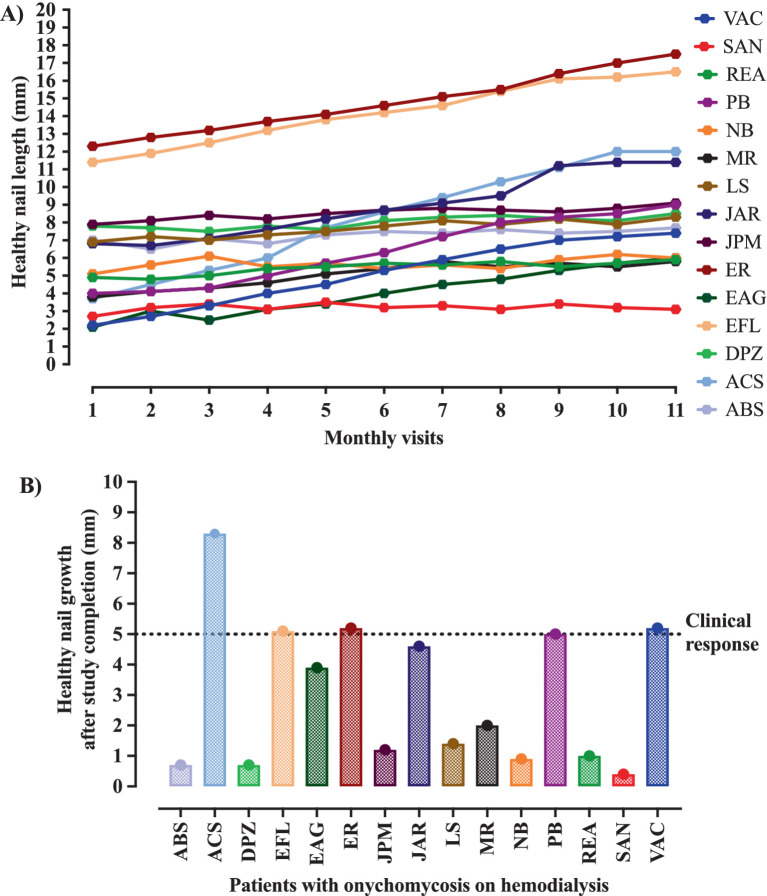
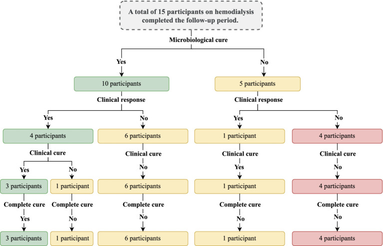
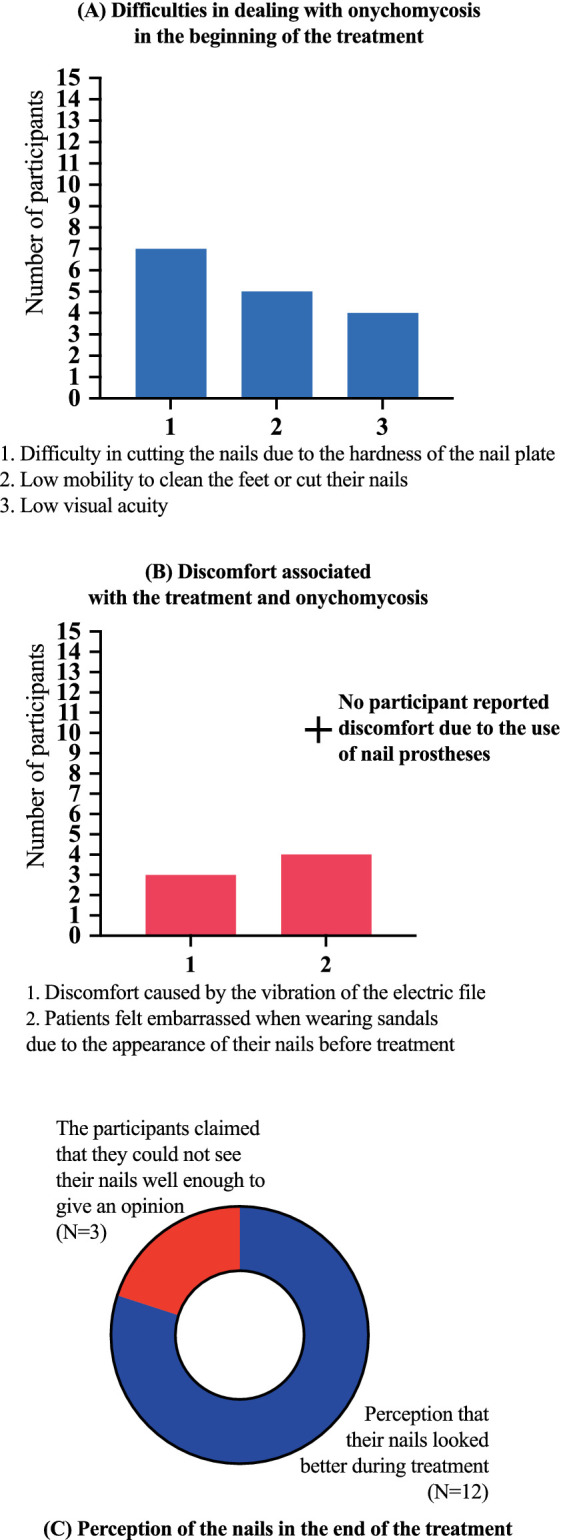
Results: Out of the 155 patients on hemodialysis, 64/155 (41.3%) individuals were identified with symptoms suggestive of onychomycosis in the halluces after clinical analysis. Among them, 35/64 (54.7%) individuals presented a positive direct mycological examination and underwent fungal culture to identify the etiological agent. In this group of patients, 24/35 (68.6%) individuals who presented clinical forms whose treatment involved the need for nail debridement were selected. Only 15/24 (62.5%) individuals completed the study. Among the study participants, 5/15 (33.3%) still presented positive fungal culture in the presence of a negative direct mycological examination and 1/15 (6.7%) presented a positive direct mycological examination, but with a negative culture. Among those with a positive fungal culture, 3/15 (20.0%) participants presented microorganisms different from those isolated in the initial exams. Regarding cure, 5/15 (33.3%) participants showed a clinical response, 4/15 (26.7%) clinical cure, and 3/15 (20.0%) complete cure. No patient presented an allergic reaction or local irritation caused by the material used in the treatment. There were accidental superficial ulcerations caused by the electric sandpaper; however, no wound developed secondary infection. No participant reported discomfort due to the nail prosthesis use, 3/15 (20.0%) reported a feeling of discomfort caused by the vibration of the electric file and 12/15 (80.0%) reported the perception that their nails had a better appearance during treatment with nail prosthesis made with acrylic reconstruction gel and 2% terbinafine.

Conclusion: The application of 2% terbinafine in acrylic reconstruction gel for the manufacture of nail prostheses applied after debridement of moderate and severe forms of onychomycosis showed low efficacy as an isolated treatment in patients on dialysis due to chronic kidney disease. On the other hand, most patients had a good perception of the appearance of their nails during treatment, even when it did not result in apparent clinical improvement or complete cure.

Keywords: antifungal; clinical trial; kidney dialysis; microbiology; nail plate debridement; onychomycosis.


Conflict of interest statement

The authors declare that the research was conducted in the absence of any commercial or financial relationships that could be construed as a potential conflict of interest.

Figures

Figure 1
Figure 1
Study protocol showing the participants’ inclusion and their follow-up during the intervention period. Intervention was carried out with debridement of the nail followed by the application of acrylic polymer with 2% terbinafine. Participants were over 18 years old and presented onychomycosis of hallux, whose treatment involved debridement of the ungual plate (dermatophytoma, +2 mm thick subungual keratosis, or prolonged onycholysis). During the study period, nine participants discontinued the follow-up, as follows: (a) four participants died due to complications of the kidney disease, (b) three participants received a transplant and stopped to use the dialysis service, and (c) two participants decided not to continue in the study for personal reasons. Cure criteria were set based on the clinical progression and laboratory exam results, and were designed as follows: (clinical response) healthy nail 5 mm growth, (mycological cure) negative direct mycological and culture, (clinical cure) completely normal nail appearance, and (complete cure) presence of clinical and mycological cure.
Figure 2
Figure 2
Evolution of the nail growth during the intervention period. The intervention was carried out with the nail debridement followed by the application of acrylic polymer with 2% terbinafine. The clinical response was positive in the presence of at least 5 mm healthy nail during the follow-up period. (A) All participants showed healthy nail growth. (B) The healthy nail growth was higher than that set as a positive response in only five cases, who showed 5-, 5.1-, 5.2-, 5.2-, and 8.3-mm nail growth, respectively, in the PB, EFL, ER, VAC, and ACS cases.
Figure 3
Figure 3
The images show two severe onychomycosis cases in which a complete cure was observed after the intervention period carried out with nail debridement followed by the application of acrylic polymer with 2% terbinafine. In both cases, we can see as follows: (A) the appearance of the hallux nail at the time of the initial evaluation – study inclusion period, when the direct mycological and fungal culture exams were carried out; (B) the appearance of the hallux nail after the first debridement and cleaning. The ungual plate debridement was carried out only in the cases indicated such as the presence of dermatophytoma, +2 mm thick subungual keratosis, or prolonged onycholysis; (C) the nail appearance during the intervention period. It seems relevant to observe that the polymerized acrylic gel with 2% terbinafine guaranteed the nail healthy appearance. (D) Nail appearance after 11 months of treatment with the polymerized acrylic gel with 2% terbinafine. Both cases resulted in a complete cure and absence of infectious agents according to the clinical and laboratory analyses.
Figure 4
Figure 4
The images show a severe onychomycosis case in which a complete cure was observed after the intervention period carried out with nail debridement followed by the application of acrylic polymer with 2% terbinafine. (A) Appearance of the nail with onychomycosis before debridement in the presence of a 35-point Onychomycosis Severity Index (OSI). (B) Nail appearance immediately after the first debridement. The OSI value reduced from 35 (time of inclusion in the study) to 25 points, due to the removal of the subungual hyperkeratosis. Thus, to evaluate the therapeutical response, the remaining ungual plate area was measured aided by the Image J, 1,47v software (NHI Bethesda, MD, EUA). During the maintenance, whenever there was healthy nail growth, the increased area was added, but when dystrophic nail growth was observed, it was debrided again and, consequently the increased area removed was not recorded. (C) Nail appearance after the intervention period with a 0-point OSI. The onychomycosis severity was assessed using the OSI system. The final score was obtained by multiplying the involvement area score (ranging from 0 to 5) by the disease proximity with the matrix score (ranging from 1 to 5). Ten points were added in the presence of subungual hyperkeratosis dermatophytoma +2 mm thick or longitudinal band. After having the scores measured, the patients subjected to hemodialysis were classified in the following severity groups: mild onychomycosis [score from 1 to 5], moderate onychomycosis [score from 6 to 15], and severe onychomycosis [score from 16 to 35] (54).
Figure 5
Figure 5
The images show a severe onychomycosis case in which a complete cure was observed after the intervention period carried out with nail debridement followed by the application of acrylic polymer with 2% terbinafine. (A) Nail appearance with mixed pattern onychomycosis, involvement of the ungual plate in the whole extension, in the presence of over 2 mm subungual hyperkeratosis. The Onychomycosis Severity Index (OSI) score was 35 points at the time of inclusion in the study. (B) Ungual plate appearance without clinical alteration after the intervention period with a 0-point OSI. The onychomycosis severity was assessed using the OSI. The final score was obtained by multiplying the involvement area score (ranging from 0 to 5) by the disease proximity with the matrix score (ranging from 1 to 5). Ten points were added in the presence of subungual hyperkeratosis dermatophytoma +2 mm thick or longitudinal band. After having the scores measured, the patients subjected to hemodialysis were classified in the following severity groups: mild onychomycosis [score from 1 to 5], moderate onychomycosis [score from 6 to 15], and severe onychomycosis [score from 16 to 35] (54).
Figure 6
Figure 6
The images show a severe onychomycosis case in which a complete cure was observed after the intervention period carried out with nail debridement followed by the application of acrylic polymer with 2% terbinafine. (A) Mixed pattern onychomycosis with 26 to 50% involvement of the ungual plate, reaching segment 3. Presence of over 2 mm subungual hyperkeratosis. The Onychomycosis Severity Index (OSI) score was 19 points at the time of inclusion in the study. (B) After the intervention period, the ungual plate did not show clinical alterations and a 0-point OSI was recorded. The onychomycosis severity was assessed using the OSI system. The final score was obtained by multiplying the involvement area score (ranging from 0 to 5) by the disease proximity with the matrix score (ranging from 1 to 5). Ten points were added in the presence of subungual hyperkeratosis dermatophytoma +2 mm thick or longitudinal band. After having the scores measured, the patients subjected to hemodialysis were classified in the following severity groups: mild onychomycosis [score from 1 to 5], moderate onychomycosis [score from 6 to 15], and severe onychomycosis [score from 16 to 35] (54).
Figure 7
Figure 7
Summary of the main findings of the study with the outcomes observed. The outcomes were evaluated after the intervention period. The intervention was carried out with the nail debridement followed by the application of acrylic polymer with 2% terbinafine. Cure criteria were set based on the clinical progression and the laboratory exams and were designed as follows: (clinical response) healthy nail 5 mm growth, (mycological cure) negative direct mycological and fungal culture exams, (clinical cure) nail with a completely normal appearance, and (complete cure) presence of clinical and mycological cure.
Figure 8
Figure 8
Results of the questionnaire about their perception of onychomycosis and the treatment development. (A) At the beginning of the treatment, 7/15 (46.7%) participants reported difficulty in cutting their nails due to the hardness of the nail plate, even with the help of family members, 5/15 (33.3%) reported that they did not have enough mobility to clean their feet or cut their nails, 4/15 (26.7%) reported that they did not see well enough and were therefore unable to clean their feet and cut their nails. (B) No participant reported discomfort due to the use of nail prostheses, 3/15 (20.0%) reported a feeling of discomfort caused by the vibration of the electric file, 4/15 (26.7%) reported that they felt embarrassed when wearing sandals due to the appearance of their nails before treatment. (C) A total of 12/15 (80.0%) participants reported the perception that their nails looked better during treatment, and 3/15 (20.0%) claimed that they could not see their nails well enough to give an opinion. No participant presented an allergic reaction or local irritation caused by the material used in the treatment. There were superficial and accidental ulcerations caused by electric sandpaper. No wound developed a secondary infection.
Figure 9
Figure 9
Identification of terbinafine in nail sample by mass spectrometry. Upper left = total ion current (TIC) of terbinafine standard. Upper right = MS2 profile of terbinafine, showing the parent ion (m/z 291) and its fragments (m/z 187). Middle left = TIC of negative control gel, showing no signal for terbinafine. Lower left = MS2 profile of nail sample containing terbinafine gel, showing the same ions as the standard.

Similar articles

References

    1. Bodman MA, Syed HA, Krishnamurthy K. Onychomycosis. Treasure Island, FL: StatPearls Publishing; (2024). - PubMed
    1. Maskan Bermudez N, Rodríguez-Tamez G, Perez S, Tosti A. Onychomycosis: old and new. J Fungi (Basel). (2023) 9:559. doi: 10.3390/jof9050559, PMID: - DOI - PMC - PubMed
    1. Bersano JMQB, Cordeiro MG, Marson FAL. Prevalence and risk predictors of onychomycosis in patients on hemodialysis: an observation, prospective, and unicenter study in Brazil. Front Med (Lausanne). (2023) 10:1268324. doi: 10.3389/fmed.2023.1268324, PMID: - DOI - PMC - PubMed
    1. Gupta AK, Stec N, Summerbell RC, Shear NH, Piguet V, Tosti A, et al. . Onychomycosis: a review. J Eur Acad Dermatol Venereol. (2020) 34:1972–90. doi: 10.1111/jdv.16394 - DOI - PubMed
    1. Gupta AK, Versteeg SG, Shear NH. Onychomycosis in the 21st century: an update on diagnosis, epidemiology, and treatment. J Cutan Med Surg. (2017) 21:525–39. doi: 10.1177/1203475417716362, PMID: - DOI - PubMed

LinkOut - more resources