. 2024 Nov 6;32(4):101372.
doi: 10.1016/j.omtm.2024.101372. eCollection 2024 Dec 12.

Exogenous expression of ATP8, a mitochondrial encoded protein, from the nucleus in vivo

Affiliations

Exogenous expression of ATP8, a mitochondrial encoded protein, from the nucleus in vivo

David V Begelman et al. Mol Ther Methods Clin Dev. .

Abstract

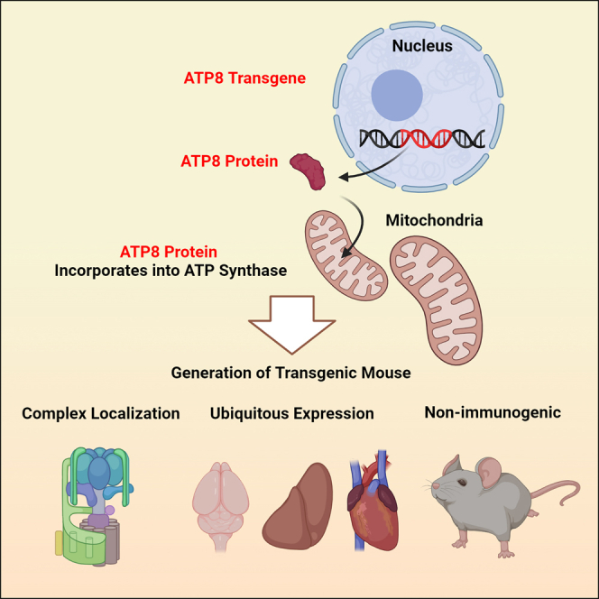
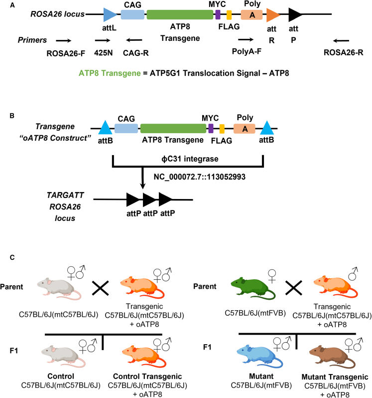
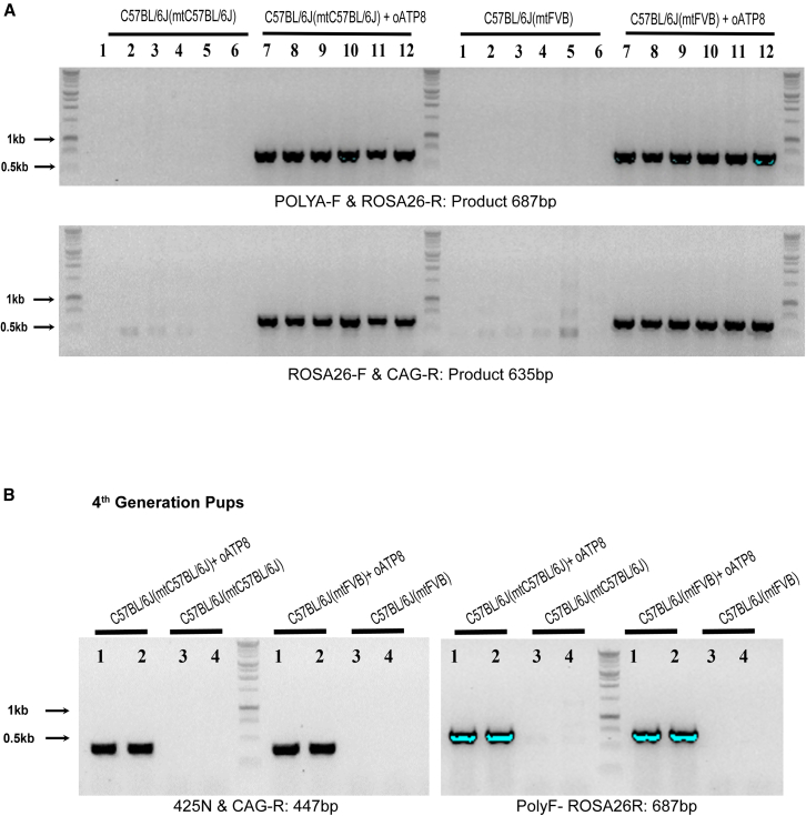
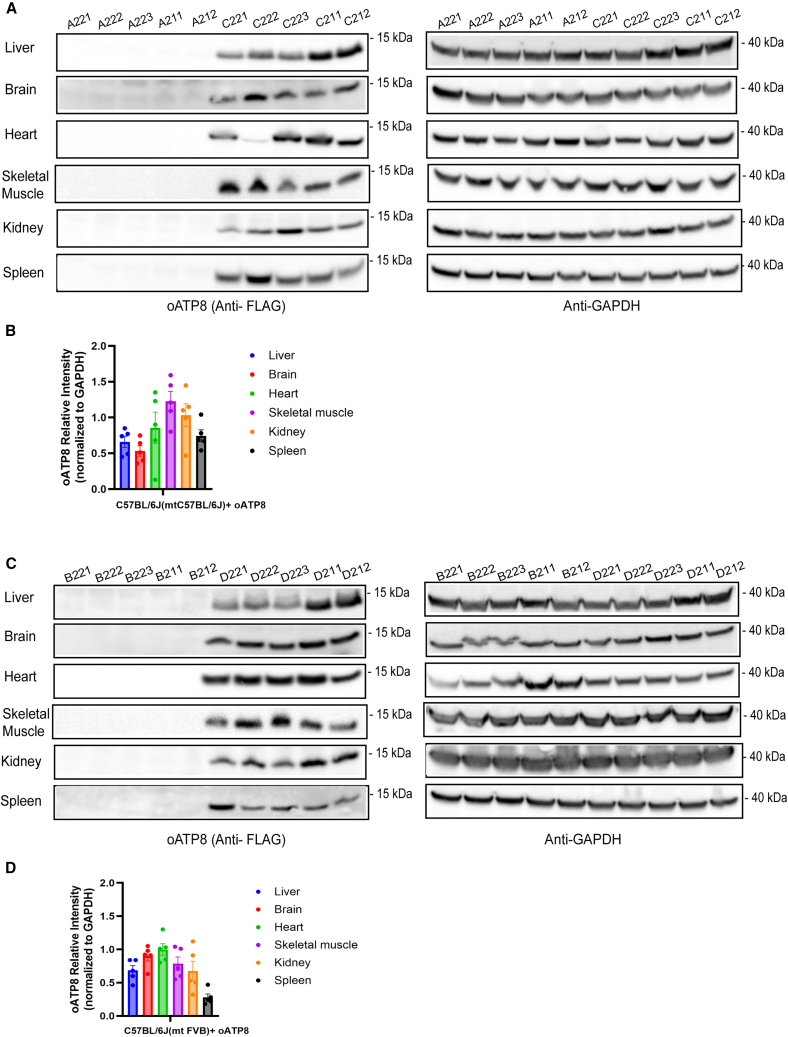
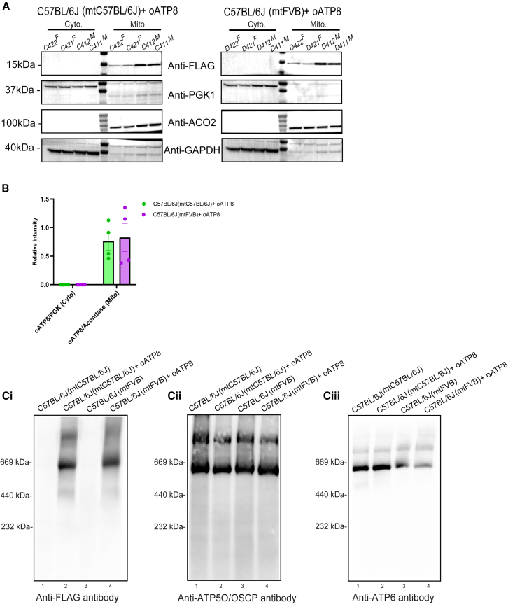
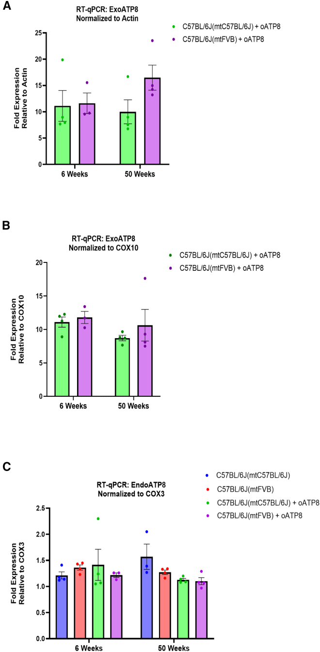
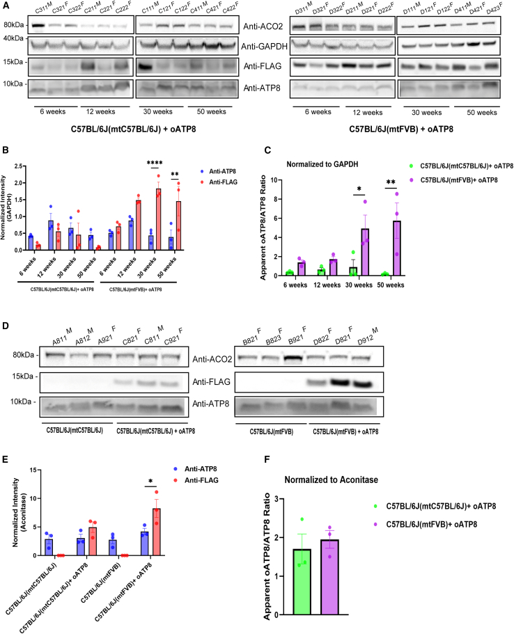
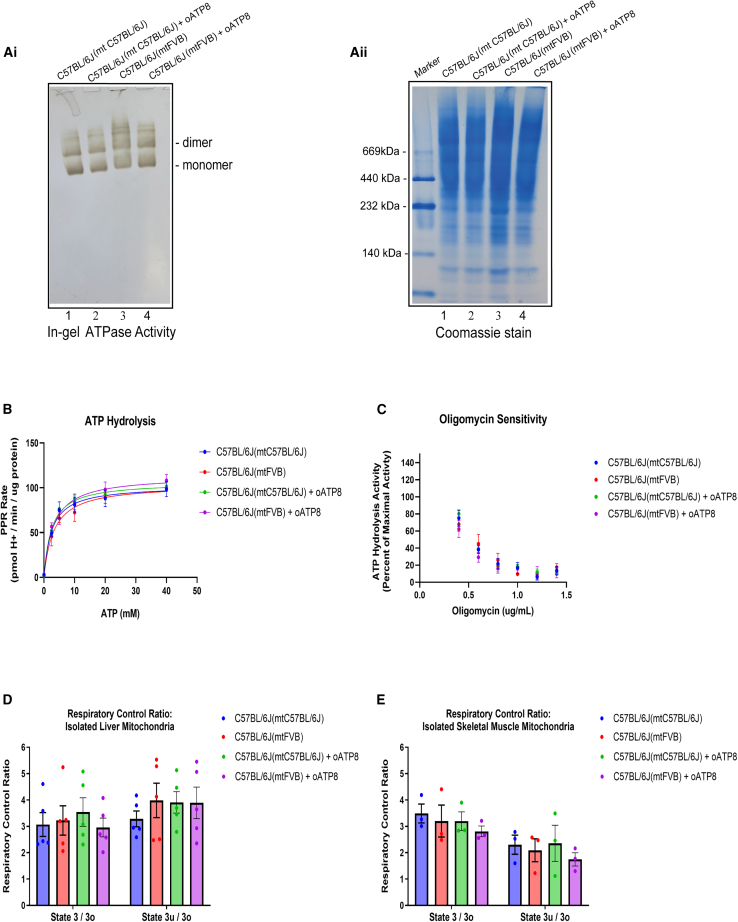
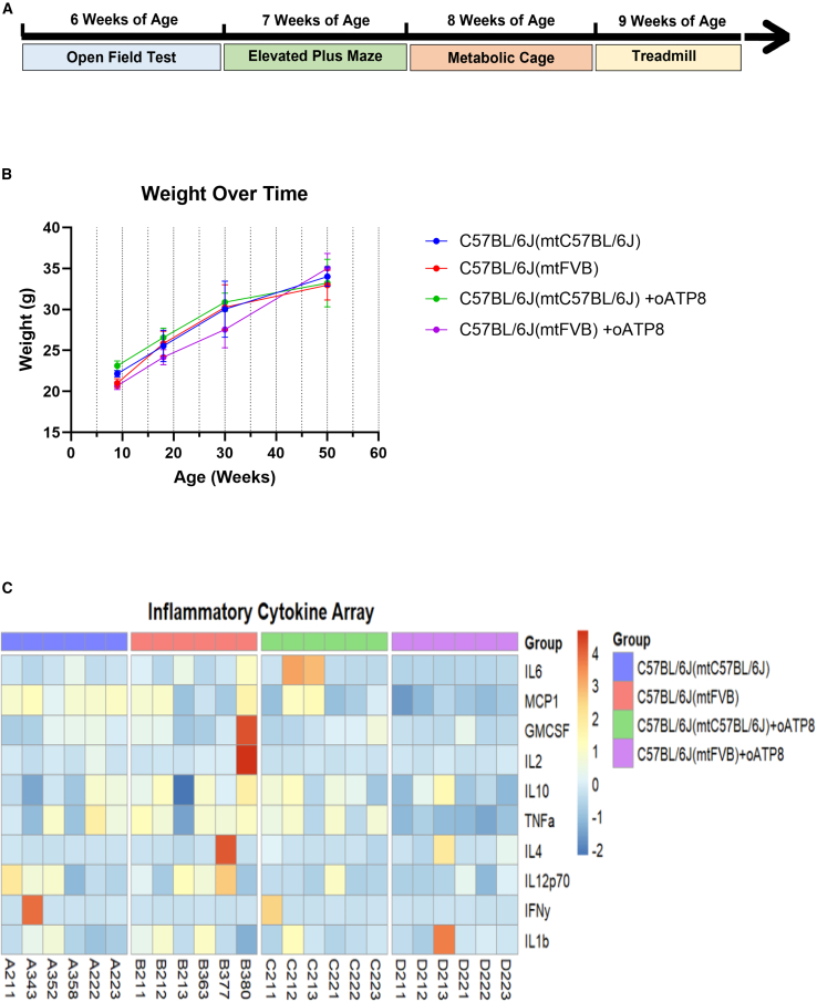
Replicative errors, inefficient repair, and proximity to sites of reactive oxygen species production make mitochondrial DNA (mtDNA) susceptible to damage with time. We explore in vivo allotopic expression (re-engineering mitochondrial genes and expressing them from the nucleus) as an approach to rescue defects arising from mtDNA mutations. We used a mouse strain C57BL/6J(mtFVB) with a natural polymorphism (m.7778 G>T) in the mitochondrial ATP8 gene that encodes a protein subunit of the ATP synthase. We generated a transgenic mouse with an epitope-tagged recoded mitochondrial-targeted ATP8 gene expressed from the ROSA26 locus in the nucleus and used the C57BL/6J(mtFVB) strain to verify successful incorporation. The allotopically expressed ATP8 protein in transgenic mice was constitutively expressed across all tested tissues, successfully transported into the mitochondria, and incorporated into ATP synthase. The ATP synthase with transgene had similar activity to non-transgenic control, suggesting successful integration and function. Exogenous ATP8 protein had no negative impact on measured mitochondrial function, metabolism, or behavior. Successful allotopic expression of a mitochondrially encoded protein in vivo in a mammal is a step toward utilizing allotopic expression as a gene therapy in humans to repair physiological consequences of mtDNA defects that may accumulate in congenital mitochondrial diseases or with age.

Keywords: ATP8 gene; allotopic expression; in vivo gene therapy; mitochondrial DNA mutation; mtDNA; safe harbor expression; transgenic mouse.


Conflict of interest statement

A patent application has been filed on the "Allotopic expression of mtDNA genes" in 2023 in the USA (PCT/US23/76302) (B.D. and A.B.). The authors declare no other competing interests.

Figures

None
Graphical abstract
Figure 1
Figure 1
Generation of transgenic mouse expressing optimized ATP8 from a safe harbor locus in the nucleus Schematic of the oATP8 construct (A) and diagram portraying the ɸC31-mediated recombination of the oATP8 construct into the mouse ROSA26 locus (NC_000072.7::113052993) (B). An overview of the breeding scheme used to generate the experimental groups (C).
Figure 2
Figure 2
Confirmation of transgene in C57BL/6J(mtC57BL/6J) and C57BL/6J(mtFVB) Sequencing products utilizing primers spanning transgene and insertion site for identification of transgene and site-specific insertion into the ROSA26 locus (n = 6 animals) (A). PCR results confirm the presence of the oATP8 construct within the fourth generation of mouse pups born from transgenic C57BL/6J(mtC57BL/6J) or C57BL/6J(mtFVB) parents (B).
Figure 3
Figure 3
Exogenous optimized ATP8 is ubiquitously expressed Denaturing PAGE western blots from processed liver, brain, heart, skeletal muscle, kidney, and spleen of 12-week-old non-transgenic and transgenic C57BL/6J(mtC57BL/6J) (A) and C57BL/6J(mtFVB) (C) mice expressing the oATP8 protein. The C-terminal FLAG epitope was immunodetected with mouse anti-FLAG antibody against the oATP8 protein. GAPDH was used as a loading control. Approximately twenty-five micrograms of protein was loaded per lane (n = 5 biological replicates). Values are expressed as means ± standard error of the mean. The immunoblot bands of oATP8 were quantified by densitometry analysis (using ImageJ), normalized to GAPDH (B and D).
Figure 4
Figure 4
Transgenic optimized ATP8 localizes to mitochondria and incorporates into ATP synthase complex Cytoplasmic (Cyto.) and mitochondrial (Mito.) fractions were isolated from liver from 50-week-old transgenic C57BL/6J(mtC57BL/6J) and C57BL/6J(mtFVB) mice. Denaturing PAGE western blots were used to assess the compartmentalization of oATP8 to the mitochondria by probing for oATP8 using mouse anti-FLAG, cytosolic contents using rabbit anti-PGK1, and mitochondrial protein using mouse anti-ACO2. GAPDH was used as a loading control. The animal ID alongside the gender (“M” for male and “F” for female) is listed at the top of the lanes. Cytoplasmic fractions were run with ∼50 μg protein and mitochondrial fractions with ∼35 μg protein per lane (n = 4 biological replicates; 2 males and 2 females) (A). The immunoblot bands of oATP8 were quantified by densitometry analysis (using ImageJ), normalized to aconitase for mitochondrial fractions, and to PGK1 for cytoplasmic fractions (B). Values are expressed as means ± standard error of the mean. Blue Native PAGE western blots using 25 μg protein from purified liver mitochondrial fractions of 12-week-old non-transgenic and transgenic C57BL/6J(mtC57BL/6J) and C57BL/6J (mtFVB mice). Integration of oATP8 into ATP synthase complex was detected by ATP synthase complex monomer (∗) and dimer (∗∗) formation using mouse anti-FLAG antibody (Ci). ATP5O/OSCP (Cii), and ATP6 (Ciii) were probed as controls for ATP synthase complex proteins (n = 3 animals).
Figure 5
Figure 5
Stable expression of transgenic oATP8 with time Quantitative RT-PCR detection of mRNA levels for transgenic oATP8 and endogenous ATP8 from brain of 50-week-old mice. Transgenic oATP8 was normalized to ACTIN (A) and COX10, a nuclear-encoded mitochondrial gene (B). Endogenous ATP8 mRNA was normalized to COX3, a mitochondria-encoded gene (C) (n = 3–4 animals, performed in triplicate). Transgenic oATP8 levels were not detected in non-transgenic animals (data not shown). Error bars show SEM. two-way ANOVA was performed.
Figure 6
Figure 6
Distribution of transgenic versus endogenous ATP8 Denaturing PAGE western blots of mouse liver tissue from 6-, 12-, 30-, and 50-week-old transgenic C57BL/6J(mtC57BL/6J) mice (A left panel) and C57BL/6J(mtFVB) mice (A, right panel). The immunoblot bands of oATP8 and endogenous ATP8 were quantified by densitometry analysis (using ImageJ), normalized to GAPDH (B) and the ratio of oATP8 and endogenous ATP8 were calculated (C). Denaturing PAGE western blots of mouse liver mitochondria from non-transgenic and transgenic C57BL/6J(mtC57BL/6J) and C57BL/6J(mtFVB) mice (D). The immunoblot bands of oATP8 and endogenous ATP8 were quantified by densitometry analysis (using ImageJ), normalized to aconitase (E) and the ratio of oATP8 and endogenous ATP8 were calculated (F). Approximately fifty micrograms of protein was run per lane (n = 3 animals per group). The animal ID alongside the gender (“M” for male and “F” for female) is listed at the top of the lanes. FLAG-tagged oATP8 protein was immunodetected with mouse anti-FLAG antibody and endogenous ATP8 was immunodetected with rabbit anti-ATP8 antibody. GAPDH and ACO2 were used as loading controls for nuclear and mitochondrial fractions. Error bars show SEM. (B) Two-way ANOVA with Šídák’s multiple comparisons: p > 0.05, ∗∗p = 0.0018, ∗∗∗∗p ≤ 0.0001. (C) Two-way ANOVA with Šídák’s multiple comparisons: p > 0.05, ∗p = 0.0205, ∗∗p = 0.00015. (E) Two-way ANOVA with Šídák’s multiple comparisons: p > 0.05, ∗p = 0.0114. (F) Unpaired t test with Welch’s correction. p > 0.05; NS, not significant.
Figure 7
Figure 7
Exogenous expression of oATP8 protein does not negatively impact mitochondrial function In gel activity analysis in 12-week-old non-transgenic and transgenic C57BL/6J(mtC57BL/6J) and C57BL/6J(mtFVB) mice to detect ATPase activity. Mouse liver mitochondria were purified and ∼25 μg mitochondrial protein was used for clear native-PAGE. ATPase activity was documented and stained with Coomassie to detect ATP synthase monomer and dimer formation (Ai and Aii). Quantitative assessment of ATP hydrolysis activity was performed using isolated liver mitochondria from 12-week-old male mice (B). The Vmax and Km of ATP synthase to ATP substrate was calculated with the 95% confidence interval (CI). Lines were fit using “Non-linear fit: Michaelis-Menten” in GraphPad Prism. Values are means ± SEM (n = 5 animals) (Table S1). ATP synthesis and maximal respiratory capacity were monitored in isolated liver and skeletal muscle mitochondria from 9- to 12-week-old male mice. Error bars show SEM. Two-way ANOVA was performed with no significant differences between groups (n = 5 for liver mitochondria, n = 3 for skeletal muscle mitochondria) (D and E). The sensitivity of the ATP synthase to oligomycin was assessed by monitoring ATP hydrolysis activity. Values are means ± SEM (n = 3 animals) (C). Lines were fit using “Non-linear fit: [Inhibitor] vs. normalized response – Variable slope” in GraphPad Prism and IC50 values and 95% CI were calculated (Table S2).
Figure 8
Figure 8
Influence of exogenous oATP8 on physiology and behavior Schematic representing the timeline of behavioral assays conducted (A). Animal weight was monitored at 9 weeks (n = 8 animals), 18 weeks (n = 7–14 animals), 30 weeks (n = 4 animals), and 50 weeks (n = 6–8 animals) (B). Heatmap portraying the ELISA results of a pro-inflammatory cytokine assay conducted on plasma from 12-week-old mice (C).

Similar articles

References

    1. Elliott H.R., Samuels D.C., Eden J.A., Relton C.L., Chinnery P.F. Pathogenic mitochondrial DNA mutations are common in the general population. Am. J. Hum. Genet. 2008;83:254–260. doi: 10.1016/j.ajhg.2008.07.004. - DOI - PMC - PubMed
    1. Cwerman-Thibault H., Sahel J.A., Corral-Debrinski M. Mitochondrial medicine: to a new era of gene therapy for mitochondrial DNA mutations. J. Inherit. Metab. Dis. 2011;34:327–344. doi: 10.1007/s10545-010-9131-5. - DOI - PubMed
    1. Howell N., Bindoff L.A., McCullough D.A., Kubacka I., Poulton J., Mackey D., Taylor L., Turnbull D.M. Leber hereditary optic neuropathy: identification of the same mitochondrial ND1 mutation in six pedigrees. Am. J. Hum. Genet. 1991;49:939–950. - PMC - PubMed
    1. Wallace D.C., Singh G., Lott M.T., Hodge J.A., Schurr T.G., Lezza A.M., Elsas L.J., 2nd, Nikoskelainen E.K. Mitochondrial DNA mutation associated with Leber's hereditary optic neuropathy. Science (New York, N.Y.) 1988;242:1427–1430. doi: 10.1126/science.3201231. - DOI - PubMed
    1. Gerbitz K.D., Obermaier-Kusser B., Lestienne P., Zierz S., Müller-Höcker J., Pongratz D., Paetzke-Brunner I., Deufel T. Mutations of the mitochondrial DNA: the contribution of DNA techniques to the diagnosis of mitochondrial encephalomyopathies. J. Clin. Chem. Clin. Biochem. 1990;28:241–250. doi: 10.1515/cclm.1990.28.4.241. - DOI - PubMed

LinkOut - more resources