

A unique tripartite collision tumor of the esophagus: a case report and literature review
- PMID: 39659797
- PMCID: PMC11628350
- DOI: 10.3389/fonc.2024.1497154
A unique tripartite collision tumor of the esophagus: a case report and literature review
Abstract
Background: The coexistence of two or more distinct neoplasms within the same anatomical site characterizes collision tumors. While the presence of dual tumors is frequently observed in esophageal cases, the simultaneous occurrence of three distinct tumor types is extremely rare, posing significant challenges for pathological evaluation and diagnosis. Surgical resection remains the primary treatment, with generally favorable outcomes.
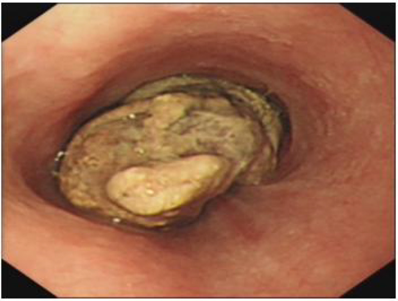
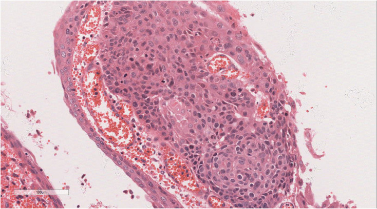
Case presentation: A 58-year-old male with a two-month history of progressively worsening dysphagia over the past 10 days underwent a gastrointestinal barium meal examination, which revealed an irregular filling defect measuring approximately 89×50 mm in the mid-thoracic esophagus. Subsequent gastroscopic biopsy confirmed undifferentiated pleomorphic sarcoma in the mid-esophageal tissue. As the dysphagia advanced, a partial esophagectomy with lymph node dissection was performed. Postoperative pathology revealed a composite tumor consisting of adenoid cystic carcinoma, undifferentiated pleomorphic sarcoma, and focal squamous cell carcinoma. Squamous cell carcinoma metastasis was identified in one lymph node. No adjuvant therapies, such as chemotherapy, radiotherapy, targeted therapy, or immunotherapy, were administered following surgery. The patient had been under monitoring for 101 months, with no signs of recurrence or metastasis.
Conclusion: This case represents the first documented instance of a tripartite collision tumor in the esophagus, composed of undifferentiated pleomorphic sarcoma, squamous cell carcinoma, and adenoid cystic carcinoma, with clear histological distinction. A thorough review of the literature was performed to summarize clinicopathological features. Surgical resection leads to a favorable prognosis. Tumors containing both carcinomatous and sarcomatous elements tend to have a more favorable prognosis compared to those composed entirely of carcinomatous tissue, providing valuable insights for future diagnostic and therapeutic strategies.
Keywords: adenoid cystic carcinoma; carcinoma; collision tumor; diagnosis; esophagus; sarcoma.
Copyright © 2024 Luo, Tian, Xu and Wang.
Conflict of interest statement
The authors declare that the research was conducted in the absence of any commercial or financial relationships that could be construed as a potential conflict of interest.
Figures


References
-
- Shi XY, Gao X, Zhong ZZ, Wang QY, Liu H. Tripartite collision tumor of the cardia: a case report and literature review. Chin J Clin Exp Pathol. (2020) 36:469–71. doi: 10.13315/j.cnki.cjcep.2020.04.024 - DOI
-
- Min Z, Qu C, Zhao Y. Esophageal collision tumor consisting of adenoid cystic carcinoma with sebaceous differentiation and squamous cell carcinoma: a case report and literature review. J Clin Exp Pathol. (2019) 35:474–6. doi: 10.13315/j.carolcarrollnkicjcep.2019.04.025 - DOI
-
- Chen L. Esophageal collision tumor. Chin Oncol. (1999) 4):91. doi: 10.19401/j.cnki.1007-3639.1999.04.032 - DOI
Publication types
LinkOut - more resources
Full Text Sources
Research Materials
Miscellaneous