

Brain and brain blood vessels histological description in autopsies of fetuses/neonates born to mothers with hypertension during pregnancy. A case-control study
- PMID: 39659801
- PMCID: PMC11629548
- DOI: 10.1016/j.lana.2024.100955
Brain and brain blood vessels histological description in autopsies of fetuses/neonates born to mothers with hypertension during pregnancy. A case-control study
Abstract
Background: Children born to women with hypertension during pregnancy have a two to threefold increased risk of developing cognitive disorders compared to children born to women without hypertension. However, structural changes in the central nervous system of these children remain poorly understood. We aim to compare the brain histological findings from autopsies of neonates and fetuses born to women with and without hypertension during pregnancy.
Methods: This retrospective case-control study includes brain histological samples from autopsies of neonates and fetuses born to women with (n = 22) and without (n = 15) hypertension during pregnancy, obtained from biobanks associated with the University Hospital San Ignacio (HUSI), Bogotá, Colombia, between 2007 and 2022. Hypertension during pregnancy was diagnosed following American College of Obstetricians and Gynecologists (ACOG) guidelines. Matched criteria included similar maternal pre-pregnancy morbidity, gestational ages at delivery, fetal sex, and availability of similar histological samples of fetal/neonatal brains. Clinical data were recorded, and two diagnosed-blinded pathologists analyzed all slides.
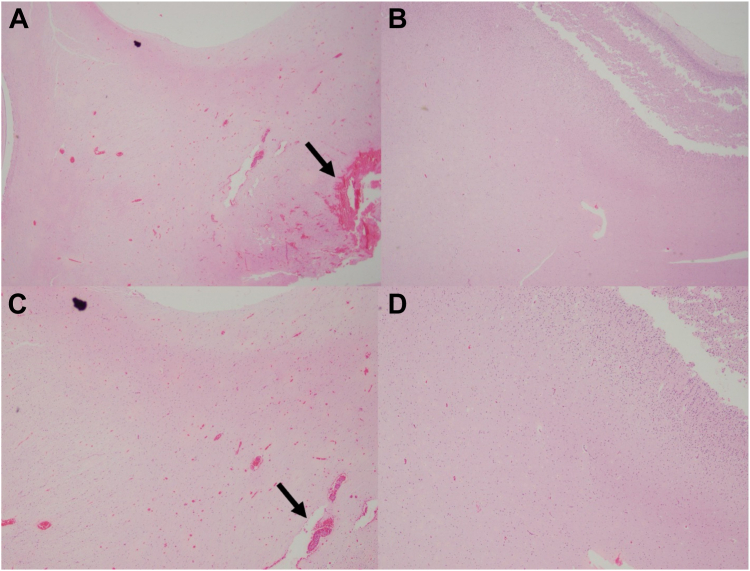
Findings: Ninety-three percent (14/15) of fetuses/neonates born to women with hypertension during pregnancy were born after preeclamptic pregnancies. Histological findings were described for the frontotemporal cortex (97%, 36/37) and meninges (81%, 30/37). Fetuses/neonates born to women with hypertension during pregnancy were smaller (p = 0.030), had a lower gestational age at death (p = 0.047), and were more frequently stillborn. Autopsy records revealed higher maternal vascular malperfusion in women with hypertension during pregnancy (p < 0.0001). Subarachnoid hemorrhage was more common in fetuses/neonates born to women with hypertension during pregnancy (p = 0.036). Other frequent findings included neuropil edema, congested meninges, hypoxic-ischemic encephalopathy, subdural hematoma, venous sinus thrombosis, hemoventricle, and necrotic foci. However, no significant endothelial or vascular wall changes were noted. "Prominent and congested" capillaries were observed only in fetuses/neonates born to women without hypertension.
Interpretation: The findings suggest increased cerebrovascular vulnerability in fetuses and neonates exposed to maternal hypertension during pregnancy, with a higher incidence of subarachnoid hemorrhage. While no vascular wall changes were identified, fewer brain capillary alterations were noted in those born to women with hypertension during pregnancy.
Funding: Fondecyt 1200250, 1240295.
Keywords: Brain; Brain angiogenesis; Fetal development; Histological evaluation; Human; Hypertensive disorders of pregnancy; Neonates; Preeclampsia.
© 2024 The Author(s).
Conflict of interest statement
The authors do not have any commercial conflicts of interest to declare. However, we initially submitted the manuscript to fulfill all the requirements for Dr. Johana Gonzalez's thesis defense as a pathologist. We used Grammarly, a typing assistant based on Artificial Intelligence, to check English texts for grammar, clarity, and engagement.
Figures
References
-
- Duley L. The global impact of pre-eclampsia and eclampsia. Semin Perinatol. 2009;33:130–137. - PubMed
-
- Narváez Diaz N. Pública Proceso de Vigilancia y Analisis de Riesgo de Salud Pública. Colombia: Instituto Nacional de Salud. FOR-R02.4000-001; 2017. Informe de evento morbilidad materna extrema, Colombia, 2017; pp. 1–17.https://www.ins.gov.co/buscador-eventos/Informesdeevento/MORBILIDAD%20MA... Accessed November 2021.
-
- ACOG, Pregnancy TFoHi. Hypertension in pregnancy. Report of the American College of Obstetricians and Gynecologists' task force on hypertension in pregnancy. Obstet Gynecol. 2013;122(5):1122–1131. - PubMed
-
- Brown M.A., Magee L.A., Kenny L.C., et al. Hypertensive disorders of pregnancy. Hypertension. 2018;72:24–43. - PubMed
LinkOut - more resources
Full Text Sources
